Zachary Lehmann
@zlehmann.bsky.social
210 followers 340 following 4 posts
Vanderbilt PhD Candidate - Cell and Developmental Biology | Tyska Lab | Microscopy, cytoskeleton, and mechanics of microscopic life 🔍
Posts Media Videos Starter Packs
Pinned
zlehmann.bsky.social
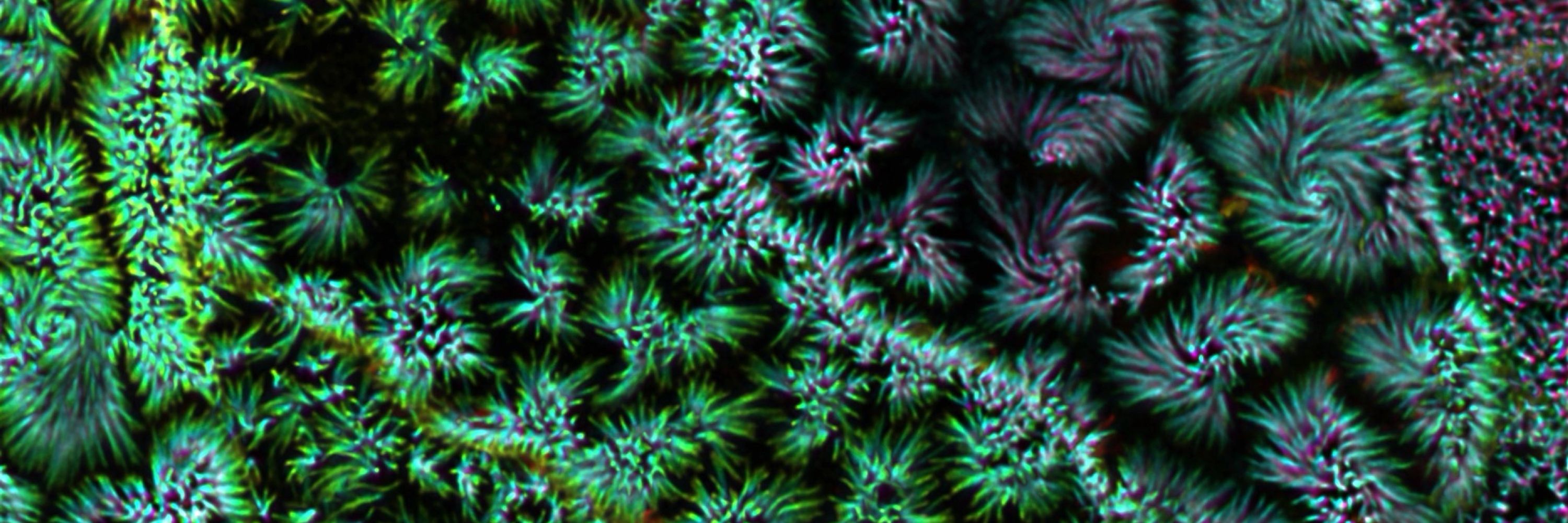
Happy #FluorescenceFriday! These are polarized epithelial cells with microvilli on their apical surface! Captured using the #Nikon NSPARC, and color-coded by depth #actin #microscopy @tyskalabactual.bsky.social
zlehmann.bsky.social
Happy #FluorescenceFriday! These are polarized epithelial cells with microvilli on their apical surface! Captured using the #Nikon NSPARC, and color-coded by depth #actin #microscopy @tyskalabactual.bsky.social
Reposted by Zachary Lehmann
tyskalabactual.bsky.social
Thought Spef1 one was some ciliary protein? Check out Dr. Rocio‘s (@rtpastrana.bsky.social) poster on how Spef1 shapes epithelial monolayers, no cilia needed 🤩. Tuesday at #cellbio2024, P2439, B46. @vucellimaging.bsky.social
Reposted by Zachary Lehmann
operkins.bsky.social
Have you always wondered how microvilli form? Where DOES that actin come from to form the core bundle? Come to the #BuildingTheCell (Part 1) subgroup tomorrow morning at 8:45 to find out! Room 29C. #CellBio2025

@tyskalabactual.bsky.social
Reposted by Zachary Lehmann
tyskalabactual.bsky.social
Also, also on Monday at #cellbio2024 I get to present Olivia’s (@operkins.bsky.social) unexpected discoveries on the link between clathrin-mediated endocytosis and the growth of epithelial microvilli 🤯
P1831. B137.
@vucellimaging.bsky.social
Reposted by Zachary Lehmann
tyskalabactual.bsky.social
Also Monday at #cellbio2024! Julissa (@yuyi106.bsky.social) will present her studies on the role of an I-BAR protein (IRTKS) in the pathogenic mechanism of Enterohemorrhagic E. coli 🦠🧫🔬 P2352. B677. @vucellimaging.bsky.social
Reposted by Zachary Lehmann
tyskalabactual.bsky.social
Posters on tuft cells are about as rare as tuft cells, but at #cellbio2024 on Monday, @jen-silverman.bsky.social presents “P1718. Organization of a cytoskeletal superstructure in the apical domain of intestinal tuft cells. B23” Yes! those are gigantic actin bundles 🤯
@vucellimaging.bsky.social
zlehmann.bsky.social
Colorful projections of filopodial tips imaged with TIRF microscopy for #FluorescenceFriday @tyskalabactual.bsky.social
Reposted by Zachary Lehmann
tyskalabactual.bsky.social
Good sampling on this confocal volume of small intestinal tissue showing the apical shag carpet (blue is F-actin, magenta is ZO1)🔬🤩
@vucellimaging.bsky.social
#microscopy #cytoskeleton #epithelial #cellbiology #biology
Reposted by Zachary Lehmann
kiannarobinson.bsky.social
It's #FluorescenceFriday fresh off the press is this depth coded image of #Microtubules. @tyskalabactual.bsky.social
Reposted by Zachary Lehmann
operkins.bsky.social
Admittedly, this is a reversed movie of #microvilli collapsing, but how cool would it be if growth were synchronized like this? #FluorescenceFriday
@tyskalabactual.bsky.social
zlehmann.bsky.social
First #FluorescenceFriday here on 🦋 Hope you enjoy this cross-section of the intestinal epithelium, highlighting the actin within the #brushborder @tyskalabactual.bsky.social
Reposted by Zachary Lehmann
operkins.bsky.social
I'm excited to rebuild this #science community over here on Bluesky! In honor of this clean slate, here's a repost of my favorite #epithelial cell building its brush border. @tyskalabactual.bsky.social
Reposted by Zachary Lehmann
tyskalabactual.bsky.social
Slicing through an intestinal organoid on the spinning disk, these cells are really dynamic 🏎️ before tissue polarity sets in.
@vucellimaging.bsky.social
zlehmann.bsky.social
Inaugural post 🦋 The apical microvilli of intestinal epithelial cells create dazzling displays as they assemble the #brushborder! This image was acquired on the remarkable #Nikon NSPARC in @tyskalabactual.bsky.social