


Francesco Padovani
@padovaf.bsky.social
Research Scientist in Cell and Computational Biology at Helmholtz Munich | Creator of Cell-ACDC and SpotMAX | Excited about mitochondrial DNA and AI-driven bioimage analysis | Dad of 2
Reposted by Francesco Padovani
Trackastra is part of the brand-new TrackMate v8.
It is the first deep-learning-based ("AI") drop-in replacement for TrackMate's regular LAP-Track algorithm.
Tracks out of the box for many types of datasets (bacteria, fluorescent nuclei, phase contrast cell culture etc), zero parameters to tune 🤖
It is the first deep-learning-based ("AI") drop-in replacement for TrackMate's regular LAP-Track algorithm.
Tracks out of the box for many types of datasets (bacteria, fluorescent nuclei, phase contrast cell culture etc), zero parameters to tune 🤖
November 20, 2025 at 8:18 PM
Trackastra is part of the brand-new TrackMate v8.
It is the first deep-learning-based ("AI") drop-in replacement for TrackMate's regular LAP-Track algorithm.
Tracks out of the box for many types of datasets (bacteria, fluorescent nuclei, phase contrast cell culture etc), zero parameters to tune 🤖
It is the first deep-learning-based ("AI") drop-in replacement for TrackMate's regular LAP-Track algorithm.
Tracks out of the box for many types of datasets (bacteria, fluorescent nuclei, phase contrast cell culture etc), zero parameters to tune 🤖
Reposted by Francesco Padovani
MASSIVE thank you to everyone involved in this year's I2K meeting - 32 SUPER sessions from our volunteer presenters, and so many super-engaged participants.
HUGE shoutout to Aditi from @bioimagingna.bsky.social for being the organizational wizard!
See you in the recordings and at next year's!
HUGE shoutout to Aditi from @bioimagingna.bsky.social for being the organizational wizard!
See you in the recordings and at next year's!
November 20, 2025 at 5:32 PM
MASSIVE thank you to everyone involved in this year's I2K meeting - 32 SUPER sessions from our volunteer presenters, and so many super-engaged participants.
HUGE shoutout to Aditi from @bioimagingna.bsky.social for being the organizational wizard!
See you in the recordings and at next year's!
HUGE shoutout to Aditi from @bioimagingna.bsky.social for being the organizational wizard!
See you in the recordings and at next year's!
Reposted by Francesco Padovani
So awesome to have this great paper from Sam Reffsin and Sara Cherry out! In it, we use retrospective clone tracing to show that there are particular single cell states that are more susceptible to viral infection (both SARS-CoV-2 and flu)!
www.cell.com/cell/fulltex...
www.cell.com/cell/fulltex...

Single-cell susceptibility to viral infection is driven by variable cell states
Not all cells that can be infected by a virus become infected with that virus. Single-cell
clone tracing reveals intrinsic cell states with variable expression patterns that
increase susceptibility to...
www.cell.com
November 14, 2025 at 4:47 PM
So awesome to have this great paper from Sam Reffsin and Sara Cherry out! In it, we use retrospective clone tracing to show that there are particular single cell states that are more susceptible to viral infection (both SARS-CoV-2 and flu)!
www.cell.com/cell/fulltex...
www.cell.com/cell/fulltex...
Reposted by Francesco Padovani
I won't talk about NMeth @natmethods.nature.com forever (probably), but I do want to brag a bit about the November focus issue on cell segmentation and tracking, which is many ways is my last hurrah (and final editorial) for the journal. I am so proud of these papers!! www.nature.com/nmeth/volume...

November 13, 2025 at 8:49 PM
I won't talk about NMeth @natmethods.nature.com forever (probably), but I do want to brag a bit about the November focus issue on cell segmentation and tracking, which is many ways is my last hurrah (and final editorial) for the journal. I am so proud of these papers!! www.nature.com/nmeth/volume...
Reposted by Francesco Padovani
Just 4 days until the start of I2K and its 33 totally free image analysis tutorials and events! Please share with your "home networks", especially early career researchers - the videos will be amazing and high-impact no matter how many people attend live, BUT (1/x)

November 13, 2025 at 2:32 PM
Just 4 days until the start of I2K and its 33 totally free image analysis tutorials and events! Please share with your "home networks", especially early career researchers - the videos will be amazing and high-impact no matter how many people attend live, BUT (1/x)
Reposted by Francesco Padovani
Applications close tomorrow - hope to see you in January! If not, stay tuned for future editions :)
Big news! Our week-long #bioimageanalysis bootcamp is BACK! Learn how to think about bioimage analysis like the pros do, learn what corrections you should (and shouldn't!) apply to your images, learn open source tools, and more. Apply NOW at broad.io/BAB3 for our January 2026 course!

November 10, 2025 at 9:23 PM
Applications close tomorrow - hope to see you in January! If not, stay tuned for future editions :)
Reposted by Francesco Padovani
From an accidental discovery of hidden biology to a new framework to understanding and diagnosing rare disease. Thrilled to share the most recent work from our lab and the amazing Jimmy Ly.
wi.mit.edu/news/alterna...
wi.mit.edu/news/alterna...

Alternate proteins from the same gene contribute differently to health and rare disease | Whitehead Institute
Iain Cheeseman and colleagues reveal the underappreciated role of single genes producing multiple proteins in atypical presentations of rare disease, and present case studies of affected patients thro...
wi.mit.edu
November 7, 2025 at 4:14 PM
From an accidental discovery of hidden biology to a new framework to understanding and diagnosing rare disease. Thrilled to share the most recent work from our lab and the amazing Jimmy Ly.
wi.mit.edu/news/alterna...
wi.mit.edu/news/alterna...
Reposted by Francesco Padovani

Seeing our good old AURA-Net from 2021 (doi.org/10.1109/ISBI...) end up 2nd on a 2025 U-Net variants leaderboard was not on my bingo card for this year, but here we are
(source: doi.org/10.48550/arX...)
(source: doi.org/10.48550/arX...)

November 5, 2025 at 1:18 PM
Seeing our good old AURA-Net from 2021 (doi.org/10.1109/ISBI...) end up 2nd on a 2025 U-Net variants leaderboard was not on my bingo card for this year, but here we are
(source: doi.org/10.48550/arX...)
(source: doi.org/10.48550/arX...)
Reposted by Francesco Padovani
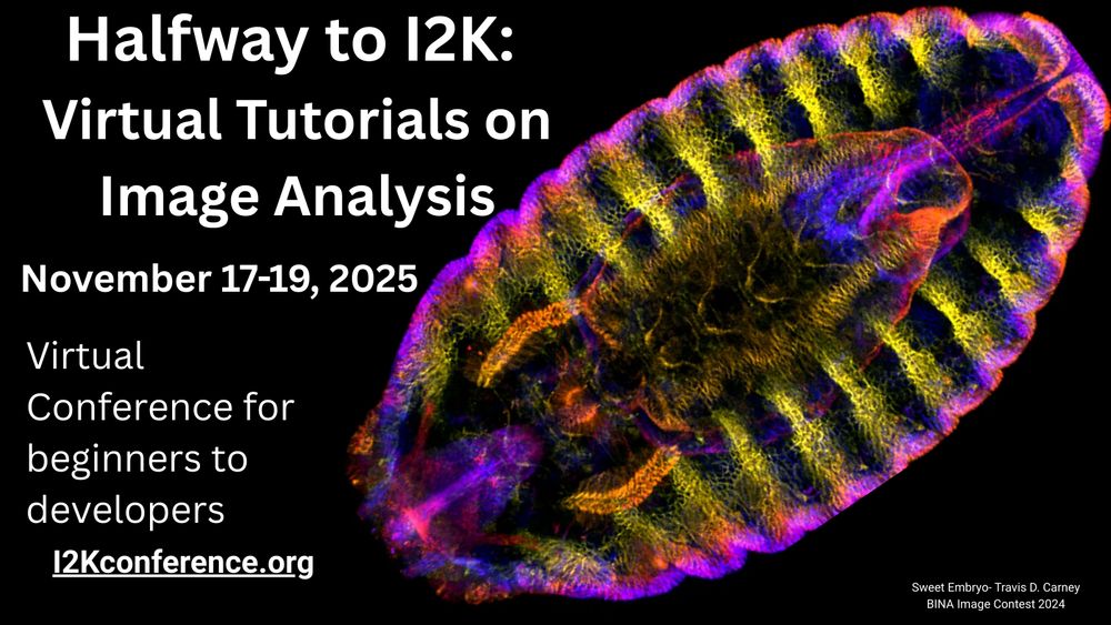
Do you want to learn more about 25+ bioimage analysis tools, 100% for free? Sign up to attend Halfway to I2K today! tinyurl.com/Halfway-to-I...
Wishing you'd submitted a tool or workshop but missed the deadline? GREAT NEWS, we just extended one week! Submit by Friday - airtable.com/app2zpB8d82r...
Wishing you'd submitted a tool or workshop but missed the deadline? GREAT NEWS, we just extended one week! Submit by Friday - airtable.com/app2zpB8d82r...

#HappyFluorescenceFriday #microscopycommunity
Got an idea for an image analysis workshop? Share it at Halfway to I2K: Online Tutorials on Image Analysis (Nov 17–19, 2025), held with Zoom Events!
Submit by Oct 31: buff.ly/lMhK4vz
Website: buff.ly/QP36RZB
Register for the workshops: buff.ly/8nNAI18
Got an idea for an image analysis workshop? Share it at Halfway to I2K: Online Tutorials on Image Analysis (Nov 17–19, 2025), held with Zoom Events!
Submit by Oct 31: buff.ly/lMhK4vz
Website: buff.ly/QP36RZB
Register for the workshops: buff.ly/8nNAI18

Airtable | Everyone's app platform
Airtable is a low-code platform for building collaborative apps. Customize your workflow, collaborate, and achieve ambitious outcomes. Get started for free.
buff.ly
November 3, 2025 at 10:04 PM
Do you want to learn more about 25+ bioimage analysis tools, 100% for free? Sign up to attend Halfway to I2K today! tinyurl.com/Halfway-to-I...
Wishing you'd submitted a tool or workshop but missed the deadline? GREAT NEWS, we just extended one week! Submit by Friday - airtable.com/app2zpB8d82r...
Wishing you'd submitted a tool or workshop but missed the deadline? GREAT NEWS, we just extended one week! Submit by Friday - airtable.com/app2zpB8d82r...
Reposted by Francesco Padovani
Registration for Halfway to I2K is now OPEN! And workshop submission is still too (for a bit)! Join 20+ presenters and counting, and hundreds of attendees in learning about image analysis, whether you're a complete beginner or a total pro.
#HappyFluorescenceFriday #microscopycommunity
Got an idea for an image analysis workshop? Share it at Halfway to I2K: Online Tutorials on Image Analysis (Nov 17–19, 2025), held with Zoom Events!
Submit by Oct 31: buff.ly/lMhK4vz
Website: buff.ly/QP36RZB
Register for the workshops: buff.ly/8nNAI18
Got an idea for an image analysis workshop? Share it at Halfway to I2K: Online Tutorials on Image Analysis (Nov 17–19, 2025), held with Zoom Events!
Submit by Oct 31: buff.ly/lMhK4vz
Website: buff.ly/QP36RZB
Register for the workshops: buff.ly/8nNAI18
Airtable | Everyone's app platform
Airtable is a low-code platform for building collaborative apps. Customize your workflow, collaborate, and achieve ambitious outcomes. Get started for free.
buff.ly
October 31, 2025 at 3:37 PM
Registration for Halfway to I2K is now OPEN! And workshop submission is still too (for a bit)! Join 20+ presenters and counting, and hundreds of attendees in learning about image analysis, whether you're a complete beginner or a total pro.
Reposted by Francesco Padovani

🔬For 22 years, we have been organizing #Mifobio "Functional Microscopy for Biology" the @cnrs.fr thematic school.
🎓 Courses, Seminars, Round Tables, Workshops. The overall theme concerns #biological_imaging in its most interdisciplinary aspects.
👉 Program: imabio-cnrs.fr
#GDRImaBio #Mifobio2025
🎓 Courses, Seminars, Round Tables, Workshops. The overall theme concerns #biological_imaging in its most interdisciplinary aspects.
👉 Program: imabio-cnrs.fr
#GDRImaBio #Mifobio2025

October 3, 2025 at 10:45 PM
🔬For 22 years, we have been organizing #Mifobio "Functional Microscopy for Biology" the @cnrs.fr thematic school.
🎓 Courses, Seminars, Round Tables, Workshops. The overall theme concerns #biological_imaging in its most interdisciplinary aspects.
👉 Program: imabio-cnrs.fr
#GDRImaBio #Mifobio2025
🎓 Courses, Seminars, Round Tables, Workshops. The overall theme concerns #biological_imaging in its most interdisciplinary aspects.
👉 Program: imabio-cnrs.fr
#GDRImaBio #Mifobio2025
Reposted by Francesco Padovani
See this? This = implanting mouse embryo. Usually this happens inside its mother and is invisible to us, but we can actually watch implantation ex vivo with the hope of understanding why implantation goes awry in embryos of older women. A 🧵...
October 1, 2025 at 6:21 PM
See this? This = implanting mouse embryo. Usually this happens inside its mother and is invisible to us, but we can actually watch implantation ex vivo with the hope of understanding why implantation goes awry in embryos of older women. A 🧵...
Reposted by Francesco Padovani
Automatic filament tracing in 3D data is now only a few clicks (or one macro) away! In the amazing #BigTrace FIJI plugin by @ekatrukha.bsky.social 🤩
My workflow will be to autotrace and then manually correct the autotraces, reducing the time for our tracing significantly!
My workflow will be to autotrace and then manually correct the autotraces, reducing the time for our tracing significantly!

New release of #BigTrace plugin is out. Together with @aafkegros.bsky.social from Simone Köhler group we added a fully automatic tracing mode. + more macro functions and bug fixes github.com/ekatrukha/Bi...
September 24, 2025 at 12:05 PM
Automatic filament tracing in 3D data is now only a few clicks (or one macro) away! In the amazing #BigTrace FIJI plugin by @ekatrukha.bsky.social 🤩
My workflow will be to autotrace and then manually correct the autotraces, reducing the time for our tracing significantly!
My workflow will be to autotrace and then manually correct the autotraces, reducing the time for our tracing significantly!
Reposted by Francesco Padovani
Halfway to I2K is BACK, friends of all kinds! Last year, 650 people attended 30+ TOTALLY FREE image analysis workshops of all kinds, across many timezones.
If you make image analysis software and want to teach it, workshop submissions are open now! We'd love to have your tool highlighted.
If you make image analysis software and want to teach it, workshop submissions are open now! We'd love to have your tool highlighted.
#HappyFluorescenceFriday!
#microscopycommunity- want to learn open source image analysis or share your knowledge to help others? We’ve got a FREE virtual workshop Nov 17-19! Now accepting workshop session applications!
Learn more & sign up: buff.ly/esGIotD
#microscopycommunity- want to learn open source image analysis or share your knowledge to help others? We’ve got a FREE virtual workshop Nov 17-19! Now accepting workshop session applications!
Learn more & sign up: buff.ly/esGIotD
September 23, 2025 at 3:48 PM
Halfway to I2K is BACK, friends of all kinds! Last year, 650 people attended 30+ TOTALLY FREE image analysis workshops of all kinds, across many timezones.
If you make image analysis software and want to teach it, workshop submissions are open now! We'd love to have your tool highlighted.
If you make image analysis software and want to teach it, workshop submissions are open now! We'd love to have your tool highlighted.
Reposted by Francesco Padovani
🚀 Our new paper is out @natmethods.nature.com!
Kuffer & Marzilli engineered conditionally stable MS2 & PP7 coat proteins (dMCP & dPCP) that degrade unless bound to RNA, enabling ultra–low-background, single-mRNA imaging in live cells.
🔗 www.nature.com/articles/s41...
🧬 www.addgene.org/John_Ngo/
Kuffer & Marzilli engineered conditionally stable MS2 & PP7 coat proteins (dMCP & dPCP) that degrade unless bound to RNA, enabling ultra–low-background, single-mRNA imaging in live cells.
🔗 www.nature.com/articles/s41...
🧬 www.addgene.org/John_Ngo/
September 22, 2025 at 6:27 PM
🚀 Our new paper is out @natmethods.nature.com!
Kuffer & Marzilli engineered conditionally stable MS2 & PP7 coat proteins (dMCP & dPCP) that degrade unless bound to RNA, enabling ultra–low-background, single-mRNA imaging in live cells.
🔗 www.nature.com/articles/s41...
🧬 www.addgene.org/John_Ngo/
Kuffer & Marzilli engineered conditionally stable MS2 & PP7 coat proteins (dMCP & dPCP) that degrade unless bound to RNA, enabling ultra–low-background, single-mRNA imaging in live cells.
🔗 www.nature.com/articles/s41...
🧬 www.addgene.org/John_Ngo/
Reposted by Francesco Padovani

It’s crucial that the bioimage community joins spaces long dominated by computer vision/CS to highlight challenges in microscopy data and how we tackle them. Brilliant talk from @aafkegros.bsky.social showing how biology is both exciting and challenging👩🏻💻🔬🧬🦠 #bioimage #microscopy #interdisciplinary
I gave a talk at Blender Conference yesterday, showing how biological volumes work, and how to visualize them in Blender 😋 youtu.be/WPajSWX730o?... #bcon25
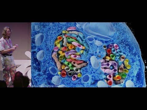
Microscopy Nodes: handling large biological volumes — Blender Conference 2025
YouTube video by Blender
youtu.be
September 19, 2025 at 8:00 AM
It’s crucial that the bioimage community joins spaces long dominated by computer vision/CS to highlight challenges in microscopy data and how we tackle them. Brilliant talk from @aafkegros.bsky.social showing how biology is both exciting and challenging👩🏻💻🔬🧬🦠 #bioimage #microscopy #interdisciplinary
Reposted by Francesco Padovani
I gave a talk at Blender Conference yesterday, showing how biological volumes work, and how to visualize them in Blender 😋 youtu.be/WPajSWX730o?... #bcon25
Microscopy Nodes: handling large biological volumes — Blender Conference 2025
YouTube video by Blender
youtu.be
September 19, 2025 at 7:35 AM
I gave a talk at Blender Conference yesterday, showing how biological volumes work, and how to visualize them in Blender 😋 youtu.be/WPajSWX730o?... #bcon25
Reposted by Francesco Padovani

No second chance for a first post - so I made it count:
Finished my PhD on glucose signaling in yeast's G2 phase.
Thanks to the whole Ewald lab and our fantastic collaborators!
Finished my PhD on glucose signaling in yeast's G2 phase.
Thanks to the whole Ewald lab and our fantastic collaborators!


July 15, 2025 at 8:43 PM
No second chance for a first post - so I made it count:
Finished my PhD on glucose signaling in yeast's G2 phase.
Thanks to the whole Ewald lab and our fantastic collaborators!
Finished my PhD on glucose signaling in yeast's G2 phase.
Thanks to the whole Ewald lab and our fantastic collaborators!
Reposted by Francesco Padovani
🔬🎤 As pre-announced a few days ago... please let us proudly present to you: 𝑴𝒊𝒄𝒓𝒐𝕊𝒑𝒍𝒊𝒕 - your ticket to imaging more, imaging more gentle, and/or imaging more efficient. 🔬
doi.org/10.1101/2025...
Like ❤️, repost 🔂, and most importantly... please send feedback ✉️ our way! 🙏
doi.org/10.1101/2025...
Like ❤️, repost 🔂, and most importantly... please send feedback ✉️ our way! 🙏

February 11, 2025 at 6:02 PM
🔬🎤 As pre-announced a few days ago... please let us proudly present to you: 𝑴𝒊𝒄𝒓𝒐𝕊𝒑𝒍𝒊𝒕 - your ticket to imaging more, imaging more gentle, and/or imaging more efficient. 🔬
doi.org/10.1101/2025...
Like ❤️, repost 🔂, and most importantly... please send feedback ✉️ our way! 🙏
doi.org/10.1101/2025...
Like ❤️, repost 🔂, and most importantly... please send feedback ✉️ our way! 🙏
Reposted by Francesco Padovani

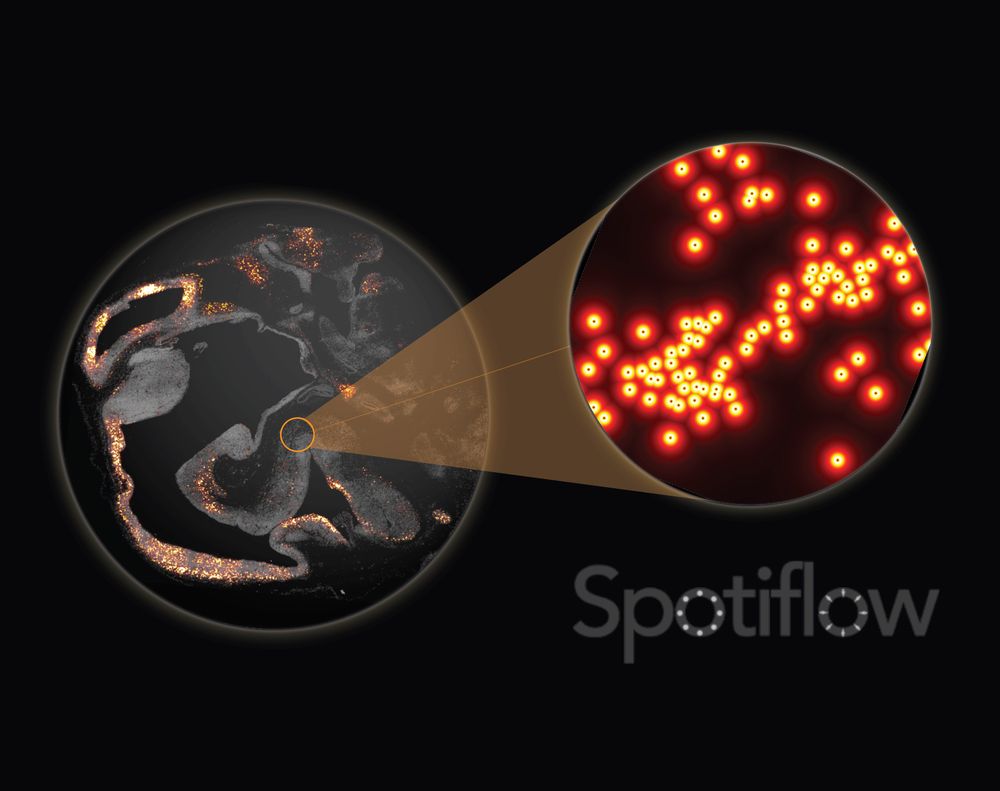
Out today in @natmethods.nature.com : Spotiflow, our transcript localization method for imaging-based spatial transcriptomics. Led by amazing PhD student @albertdm.bsky.social, joint work w @gioelelamanno.bsky.social at EPFL / @scadsai.bsky.social
www.nature.com/articles/s41...
rdcu.be/epIB7
www.nature.com/articles/s41...
rdcu.be/epIB7
June 6, 2025 at 7:06 PM
Out today in @natmethods.nature.com : Spotiflow, our transcript localization method for imaging-based spatial transcriptomics. Led by amazing PhD student @albertdm.bsky.social, joint work w @gioelelamanno.bsky.social at EPFL / @scadsai.bsky.social
www.nature.com/articles/s41...
rdcu.be/epIB7
www.nature.com/articles/s41...
rdcu.be/epIB7
Reposted by Francesco Padovani
Spotiflow, our deep learning based spot detection method for microscopy, is now published in @natmethods.nature.com!
Since the pre-print, we have added many features, notably native 3D detection!
@maweigert.bsky.social @gioelelamanno.bsky.social @epfl-brainmind.bsky.social
Paper: rdcu.be/epIB7
(1/N)
Since the pre-print, we have added many features, notably native 3D detection!
@maweigert.bsky.social @gioelelamanno.bsky.social @epfl-brainmind.bsky.social
Paper: rdcu.be/epIB7
(1/N)
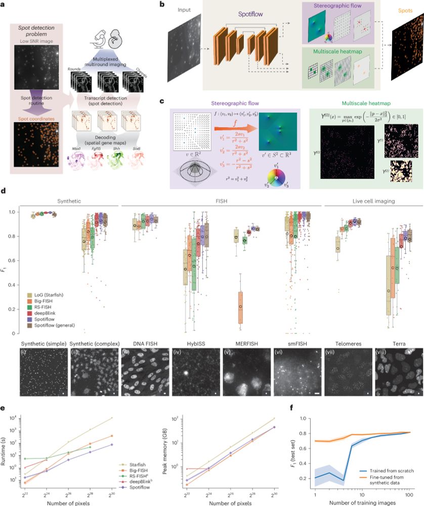
Spotiflow: accurate and efficient spot detection for fluorescence microscopy with deep stereographic flow regression
Nature Methods - Spotiflow uses deep learning for subpixel-accurate spot detection in diverse 2D and 3D images. The improved accuracy offered by Spotiflow enables improved biological insights in...
rdcu.be
June 6, 2025 at 6:58 PM
Spotiflow, our deep learning based spot detection method for microscopy, is now published in @natmethods.nature.com!
Since the pre-print, we have added many features, notably native 3D detection!
@maweigert.bsky.social @gioelelamanno.bsky.social @epfl-brainmind.bsky.social
Paper: rdcu.be/epIB7
(1/N)
Since the pre-print, we have added many features, notably native 3D detection!
@maweigert.bsky.social @gioelelamanno.bsky.social @epfl-brainmind.bsky.social
Paper: rdcu.be/epIB7
(1/N)
Reposted by Francesco Padovani
Wow, just implemented the new Cellpose-SAM from @computingnature.bsky.social in NimbusImage and it's *awesome*! Give it a try!
nimbusimage.com
nimbusimage.com

May 7, 2025 at 6:19 PM
Wow, just implemented the new Cellpose-SAM from @computingnature.bsky.social in NimbusImage and it's *awesome*! Give it a try!
nimbusimage.com
nimbusimage.com
Reposted by Francesco Padovani

⏳ Only 8 days left to register for the AI4Life Community Event in Helsinki!
Day 1️⃣: Workshops for life scientists, software devs, AI researchers & image analysts
Day 2️⃣: Talks, discussions & networking
If that sounds like you, don’t miss it!
👉 ai4life.eurobioimaging.eu/event/ai4lif...
Day 1️⃣: Workshops for life scientists, software devs, AI researchers & image analysts
Day 2️⃣: Talks, discussions & networking
If that sounds like you, don’t miss it!
👉 ai4life.eurobioimaging.eu/event/ai4lif...
April 17, 2025 at 3:25 PM
⏳ Only 8 days left to register for the AI4Life Community Event in Helsinki!
Day 1️⃣: Workshops for life scientists, software devs, AI researchers & image analysts
Day 2️⃣: Talks, discussions & networking
If that sounds like you, don’t miss it!
👉 ai4life.eurobioimaging.eu/event/ai4lif...
Day 1️⃣: Workshops for life scientists, software devs, AI researchers & image analysts
Day 2️⃣: Talks, discussions & networking
If that sounds like you, don’t miss it!
👉 ai4life.eurobioimaging.eu/event/ai4lif...
Reposted by Francesco Padovani

During the workshop earlier this week we discussed how important it is to inspect histograms of image regions in some projects. Thus, here comes stackview.histogram() - new in stackview 0.15.0 🥳
github.com/haesleinhuep...
github.com/haesleinhuep...
April 10, 2025 at 1:00 PM
During the workshop earlier this week we discussed how important it is to inspect histograms of image regions in some projects. Thus, here comes stackview.histogram() - new in stackview 0.15.0 🥳
github.com/haesleinhuep...
github.com/haesleinhuep...
Reposted by Francesco Padovani
Our latest preprint www.biorxiv.org/content/10.1..., describes Willi Stepp’s project to make smart microscopy even gentler by doing event detection in phase contrast. We developed neural networks to detect mito-LD and mito-lysosome contacts, as well as mitochondrial pre-fission constrictions.

April 6, 2025 at 11:44 AM
Our latest preprint www.biorxiv.org/content/10.1..., describes Willi Stepp’s project to make smart microscopy even gentler by doing event detection in phase contrast. We developed neural networks to detect mito-LD and mito-lysosome contacts, as well as mitochondrial pre-fission constrictions.