Patricia Ramos
@apramos.bsky.social
130 followers
250 following
15 posts
Dev Biologist, postdoc @BarrigaLab_@PolDresden. Postdoc
@NordenLab, MSCA fellow_optic cup morphogenesis, Phd @michalis_averof_parhyale eye evo-devo. #morphogenesis #evo_devo #imaging #image_analysis
Posts
Media
Videos
Starter Packs
Patricia Ramos
@apramos.bsky.social
· Sep 4
Patricia Ramos
@apramos.bsky.social
· Sep 1
Patricia Ramos
@apramos.bsky.social
· Sep 1


Patricia Ramos
@apramos.bsky.social
· Sep 1
Patricia Ramos
@apramos.bsky.social
· Sep 1
Patricia Ramos
@apramos.bsky.social
· Sep 1

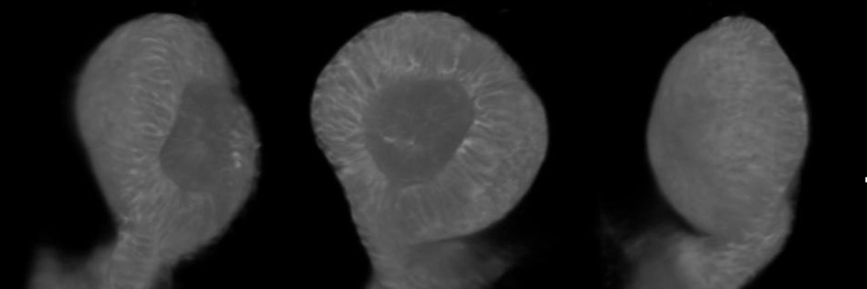
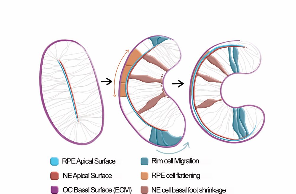
The Optic cup is actively shape programmed by independently patterned apical forces
During morphogenesis, initially flat tissues often must transition into complex 3D shapes, reminiscent of shape-programmable systems in physics and engineering. One key question in developmental biolo...
www.biorxiv.org