Manning Research Group
@manningresearch.bsky.social
150 followers 62 following 90 posts
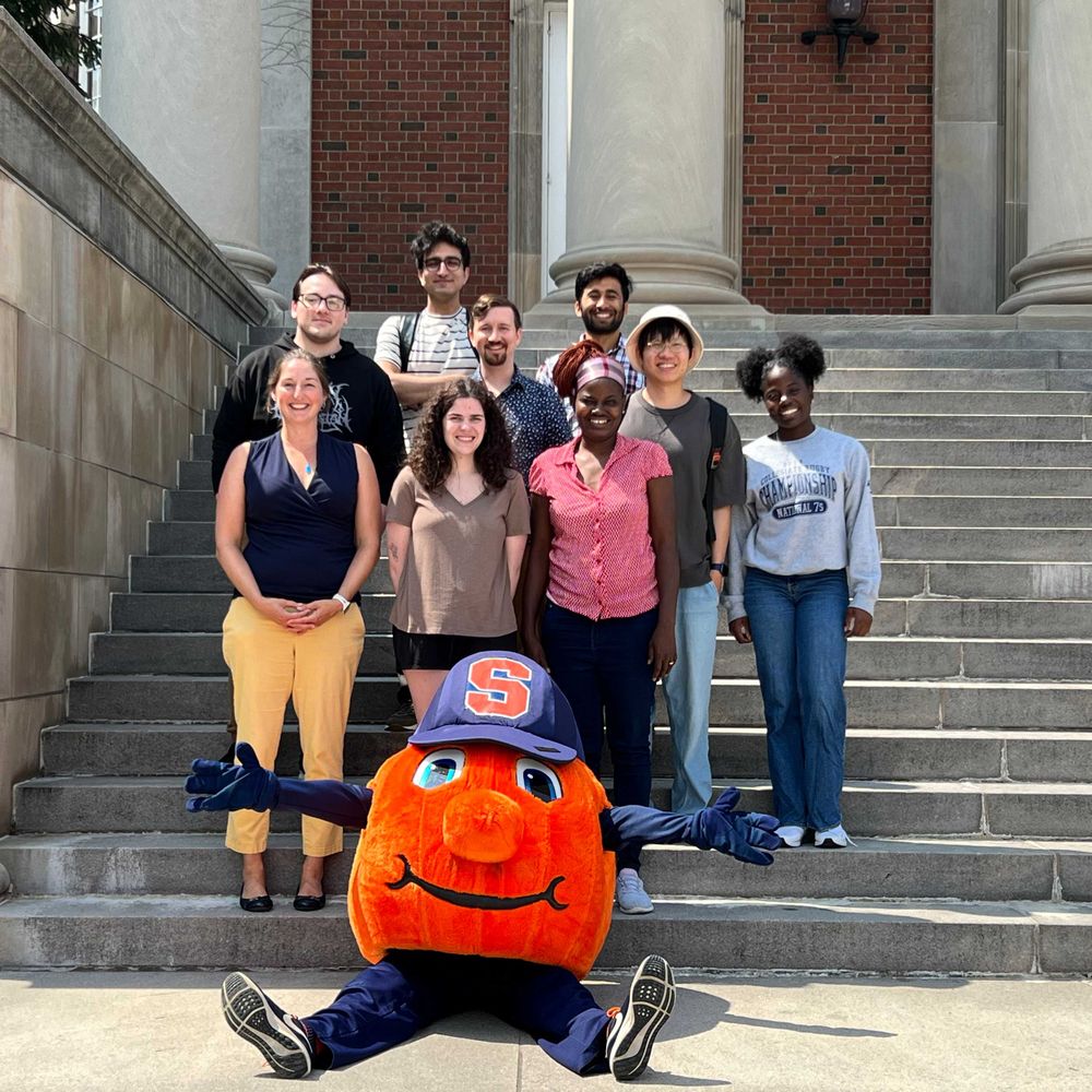
Manning Research Group at Syracuse University: theory and computation focused on cells, grains, tissues, glasses, and other out-of-equilibrium disordered matter
Posts Media Videos Starter Packs
manningresearch.bsky.social
Our work confirms that slow tissue movements can generate forces that are significant enough to deform an organ, as the timescale of tissue relaxation is large.

This suggests dynamical forces may be playing a role in many other developmental processes, too. We should look!
13/n
manningresearch.bsky.social
In addition to altering lumen shape changes, are dynamical forces sufficient to change individual cell shapes to drive KV remodeling involved in LR patterning?

Yes, notochord ablation reduces the AP distribution as compared to controls. The 3D vertex model predicts this.
12/n
manningresearch.bsky.social
These shape changes are generic – they can also be seen in a simpler hydrodynamic model of a membrane surrounded by a highly viscous medium, also with anterior pushing forces and posterior pulling forces.

11/n
manningresearch.bsky.social
The experiments match the simulation predictions! In notochord ablation experiments, the lumen elongates along the anterior-posterior axis (RgAP/RgLRincreases), while posterior cells ablations extend the lumen along the left-right axis (RgAP/RgLR decreases).

10/n
manningresearch.bsky.social
We quantify KV shape by the ratio of the radius of gyration of the lumen along the AP and LR directions (RgAP/RgLR).
In simulations, we mimic notochord (posterior cell) ablations by removing the pushing (pulling) forces, and find that the organ changes it shape significantly.
9/n
manningresearch.bsky.social
In simulations, we identify a set of model parameters (star in phase diagram) that generate the lumen shape seen in control experiments, where KV is pushed from the anterior by the notochord (orange cells) and pulled by posterior cells (purple cells).
manningresearch.bsky.social
Laser ablation of the notochord reduces the speed of KV in comparison to control experiments. That suggests that we were able to reduce the forces on the KV at the anterior part of KV.
We next ask how these reduced forces affect the shape of the KV lumen and KV cells
7/n
manningresearch.bsky.social
To test our hypothesis, we developed 3D models and laser ablation experiments to:
1) quantify how perturbing structures around KV impact its motion
2) measure cell and organ shape in these cases
3) show that observed in vivo shape changes match those predicted from the 3D model
6/n
manningresearch.bsky.social
Our hypothesis: The high-viscosity tailbud tissue exerts drag forces on KV as the organ is pushed through the tissue by convergent extension in the notochord, and pulled through the tissue by posteriorly migrating cells. These forces are sufficient to change KV shape.
5/n
manningresearch.bsky.social
To answer this question, we study Kupffer’s Vesicle (KV). KV is composed of a single layer of epithelial cells surrounding a fluid-filled lumen, located in tailbud of zebrafish, and it moves through surrounding tailbud tissue slowly, at about a micron per minute.
3/n
manningresearch.bsky.social
A key question in developmental biology is how organisms robustly control the morphology of tissues and organs. Mechanical forces can help provide such a control mechanism, but because tissue motion is so slow, most studies have assumed local force balance. Is that true?
2/n
manningresearch.bsky.social
This was a fun collaboration spearheaded by @rajphys.bsky.social and Emma Retzlaff, in collaboration with the fabulous Amack Lab and @lovelessradio.bsky.social, in the great environment of @syracuseu.bsky.social
manningresearch.bsky.social
Yay! New paper out in PNAS: www.pnas.org/doi/10.1073/... . How do dynamical forces generated by tissue movement affect organ morphology changes during embryonic development?
Using Kupffer’s vesicle in zebrafish embryo we showed that dynamical forces produce shape changes in a developing organ.
manningresearch.bsky.social
What the paper shows is that -- for some key observables -- it doesn't seem to matter which model you choose. The cell shape always predicts the rigidity transition, and the viscoelastic response is the same on either side of the transition.
manningresearch.bsky.social
But the key point is that we're not sure which of these models is "right" -- some might be right for one cell but not another. So what I mean is that different versions of vertex models are likely "wrong" for certain cell types.
manningresearch.bsky.social
One way of rationalizing the P^2 term is to say some cell types have a contractile actin ring; another way is to say that there's a limited pool in the cell of, e.g. cadherins or myosin.
manningresearch.bsky.social
The linear term is a line tension (2D version of a surface tension) but the quadratic term is more mysterious, and some other versions of vertex models remove it. Other versions of vertex models have a spring-like restoring force on each edge, instead of a restoring force on the perimeter.
manningresearch.bsky.social
We know that cell cortex mechanics is really complex (viscoelastic, with feedback loops, etc.) A standard vertex model makes a specific assumption about that mechanics -- with a term proportional to the perimeter P and a second term proportional to P^2,
manningresearch.bsky.social
Excited to have a discussion on this topic with outstanding colleagues!
cellpress.bsky.social
What happens when physics meets biology?
Dive into active matter, the driving force behind cytoskeletal organization, tissue morphogenesis & more. July 11 @ 9 a.m. ET.
http://dlvr.it/TLZGBg
Webinar
manningresearch.bsky.social
Congratulations to graduate student Tyler Hain for his paper accepted to Phys. Rev. E. Here's a link to the tweetorial (on x until we finish migration to bluesky): x.com/ManningResea...
x.com
x.com
manningresearch.bsky.social
Fun article in Washington post for kids about Bio-inspired science and engineering at Syracuse University!

x.com/SUBioInspired/…