
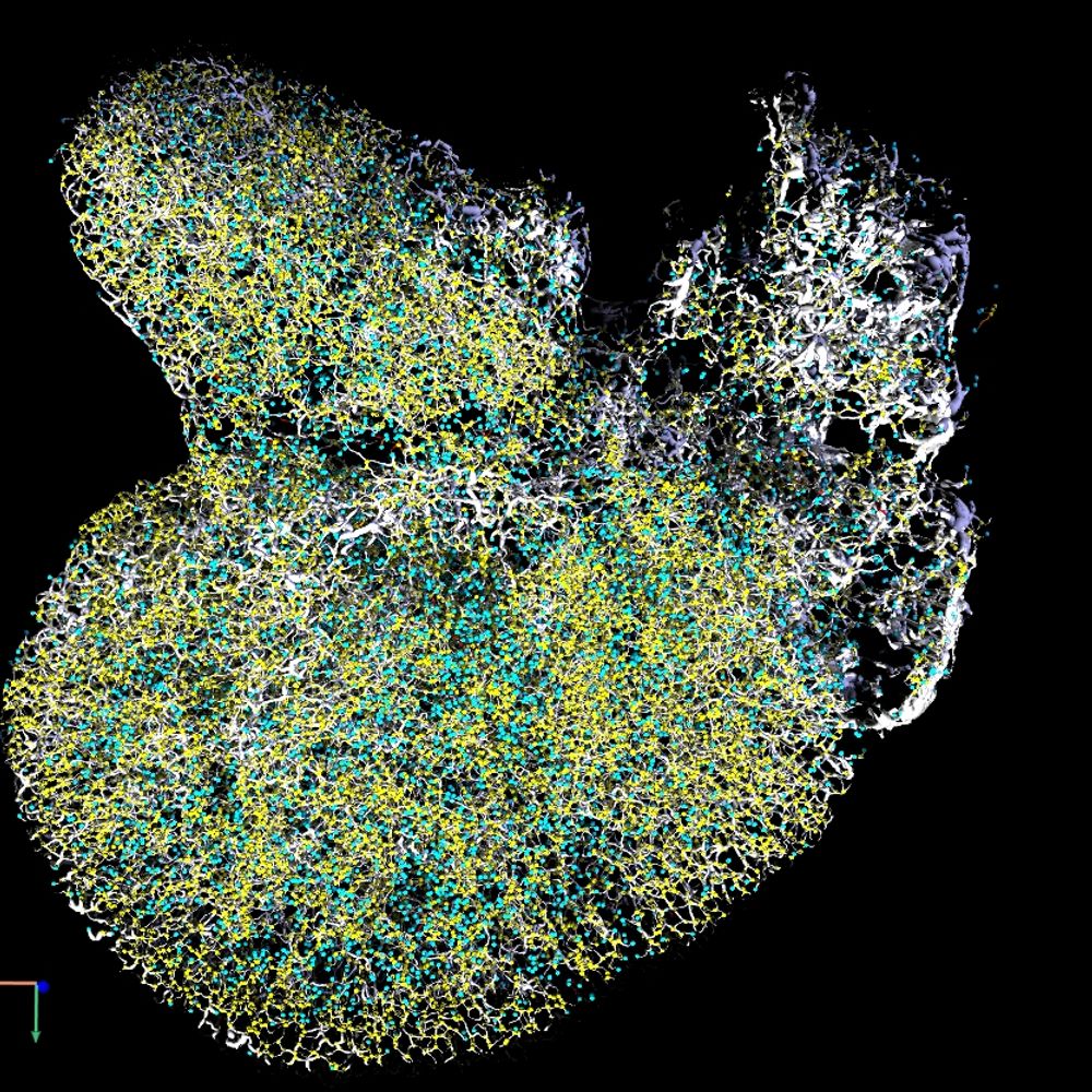
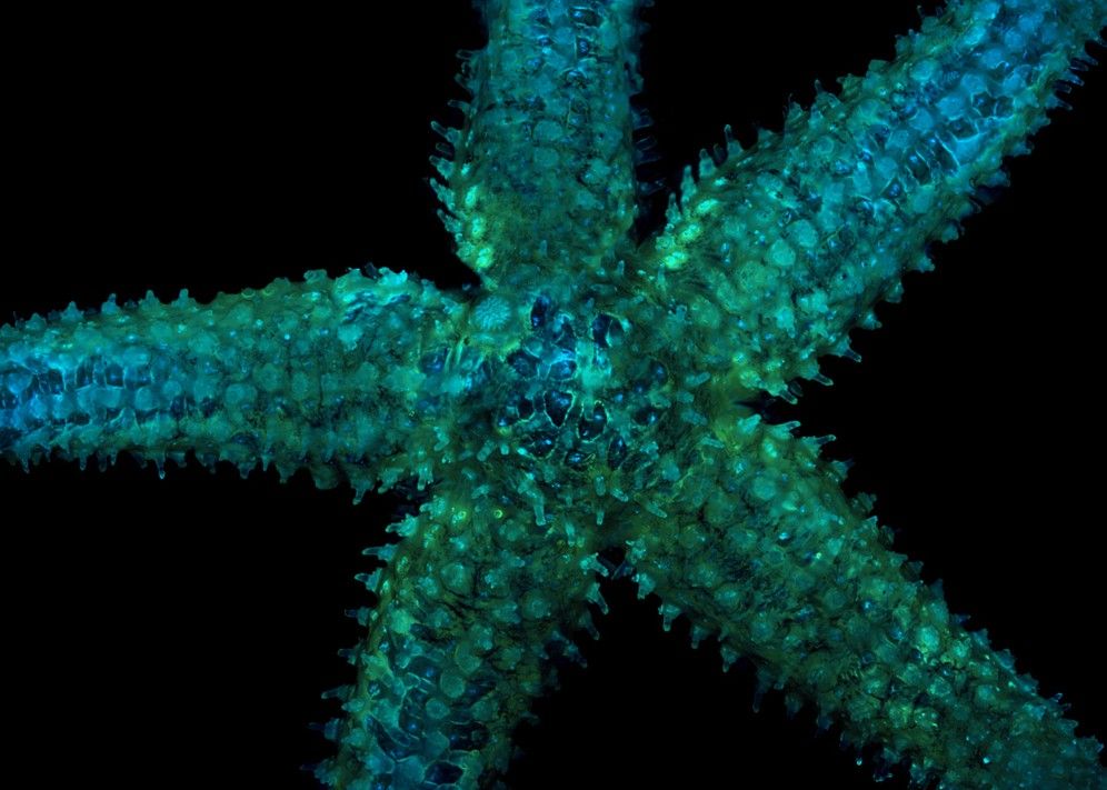
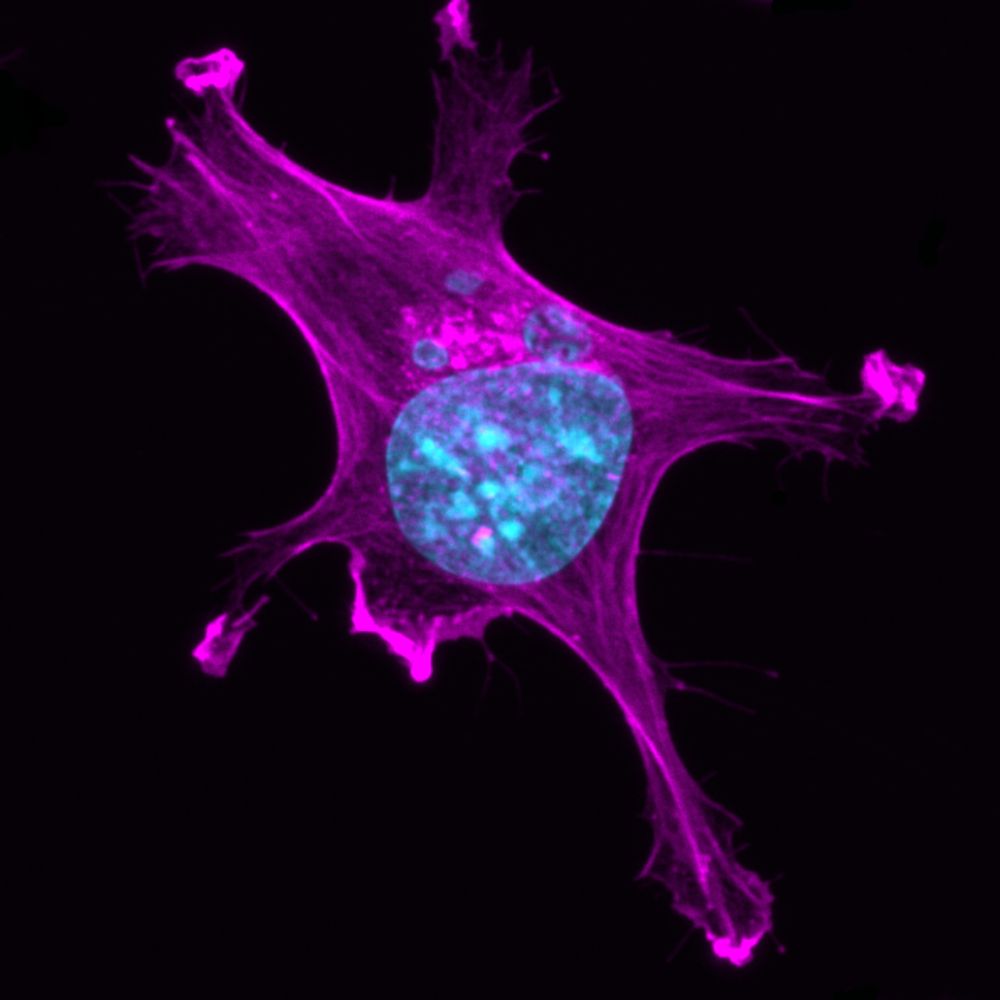
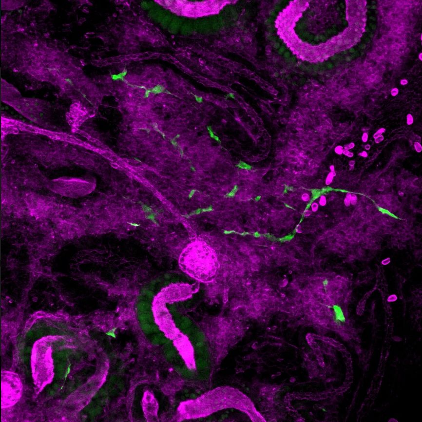
MDIBL Light Microscopy Facility
@mdibl-lmf.bsky.social
88 followers
140 following
7 posts
Light Microscopy Facility @mdibiolab.bsky.social
https://lmf.mdibl.org/
Posts
Media
Videos
Starter Packs
Reposted by MDIBL Light Microscopy Facility

Reposted by MDIBL Light Microscopy Facility


Reposted by MDIBL Light Microscopy Facility

Reposted by MDIBL Light Microscopy Facility


Reposted by MDIBL Light Microscopy Facility
Reposted by MDIBL Light Microscopy Facility
Reposted by MDIBL Light Microscopy Facility
Reposted by MDIBL Light Microscopy Facility

Reposted by MDIBL Light Microscopy Facility


Reposted by MDIBL Light Microscopy Facility

Reposted by MDIBL Light Microscopy Facility

Reposted by MDIBL Light Microscopy Facility

Reposted by MDIBL Light Microscopy Facility
Reposted by MDIBL Light Microscopy Facility


Reposted by MDIBL Light Microscopy Facility
Reposted by MDIBL Light Microscopy Facility

Reposted by MDIBL Light Microscopy Facility

