


Monique Honsa
@moniquehonsa.bsky.social
Physicist - Doctoral Candidate @jungmannlab.bsky.social
Exploring the cellular world at the nanometer scale with DNA-PAINT.
@mpibiochem.bsky.social
@lmumuenchen.bsky.social
Exploring the cellular world at the nanometer scale with DNA-PAINT.
@mpibiochem.bsky.social
@lmumuenchen.bsky.social
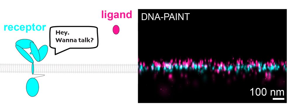
IMAGING LIGAND-RECEPTOR INTERACTIONS AT SINGLE-PROTEIN RESOLUTION WITH DNA-PAINT🔬
Ever wonder how cells "talk"? It starts when ligands bind to receptors on cell surfaces. We have cracked the challenge of imaging small ligands on cell surfaces. #DNAPAINT #celltalk 💬
doi.org/10.1002/smtd...
Ever wonder how cells "talk"? It starts when ligands bind to receptors on cell surfaces. We have cracked the challenge of imaging small ligands on cell surfaces. #DNAPAINT #celltalk 💬
doi.org/10.1002/smtd...
April 10, 2025 at 11:40 AM
IMAGING LIGAND-RECEPTOR INTERACTIONS AT SINGLE-PROTEIN RESOLUTION WITH DNA-PAINT🔬
Ever wonder how cells "talk"? It starts when ligands bind to receptors on cell surfaces. We have cracked the challenge of imaging small ligands on cell surfaces. #DNAPAINT #celltalk 💬
doi.org/10.1002/smtd...
Ever wonder how cells "talk"? It starts when ligands bind to receptors on cell surfaces. We have cracked the challenge of imaging small ligands on cell surfaces. #DNAPAINT #celltalk 💬
doi.org/10.1002/smtd...