


Zoe Kulik
@paleozoek.bsky.social
Vertebrate paleobiologist, Gerstner Scholar at the American Museum of Natural History, UW Biology alumn, associate editor @ JVP
I study the evolution of mammalian biology from extinct stem mammal fossils
🦴🩻🪚🔬👀
I study the evolution of mammalian biology from extinct stem mammal fossils
🦴🩻🪚🔬👀
Love dinosaurs? Come chat with past AMNH REU intern Jean at his poster # 539 on the second floor of Hall 3 today! #SVP2025 #2025SVP #dinosaurs #bonehistology

November 15, 2025 at 1:27 PM
Love dinosaurs? Come chat with past AMNH REU intern Jean at his poster # 539 on the second floor of Hall 3 today! #SVP2025 #2025SVP #dinosaurs #bonehistology
Reposted by Zoe Kulik
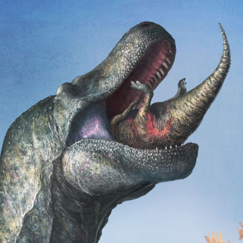
The reality of being a freelancer is that you end up working a lot of evenings and nights, something #mammals have been doing since the Mesozoic. Here's new #paleoart of two #Jurassic gliding Mammaliaformes, Arboroharamiya, starting their day as the sun sets 160 million years ago. #sciart #fossil

November 4, 2025 at 9:20 PM
Reposted by Zoe Kulik

🚨New paper alert!🚨
🧪⚒️
Terrestrial gigantism evolved through divergent pathways, with dinosaurs favoring cortical reinforcement while mammals adopted flexible trabecular expansion strategies
www.sciencedirect.com/science/arti...
🧪⚒️
Terrestrial gigantism evolved through divergent pathways, with dinosaurs favoring cortical reinforcement while mammals adopted flexible trabecular expansion strategies
www.sciencedirect.com/science/arti...

November 3, 2025 at 10:07 PM
🚨New paper alert!🚨
🧪⚒️
Terrestrial gigantism evolved through divergent pathways, with dinosaurs favoring cortical reinforcement while mammals adopted flexible trabecular expansion strategies
www.sciencedirect.com/science/arti...
🧪⚒️
Terrestrial gigantism evolved through divergent pathways, with dinosaurs favoring cortical reinforcement while mammals adopted flexible trabecular expansion strategies
www.sciencedirect.com/science/arti...
🚨 New 🦎 🚨
Breugnathair (“Bree-ack na-haird”) elgolensis is the oldest and most complete squamate from the Middle Jurassic of Scotland. Its Gaelic name means ‘false snake of Elgol’, referencing the snake-like teeth and jaws but lizard-like body and limb proportions.
Breugnathair (“Bree-ack na-haird”) elgolensis is the oldest and most complete squamate from the Middle Jurassic of Scotland. Its Gaelic name means ‘false snake of Elgol’, referencing the snake-like teeth and jaws but lizard-like body and limb proportions.


October 3, 2025 at 10:49 AM
🚨 New 🦎 🚨
Breugnathair (“Bree-ack na-haird”) elgolensis is the oldest and most complete squamate from the Middle Jurassic of Scotland. Its Gaelic name means ‘false snake of Elgol’, referencing the snake-like teeth and jaws but lizard-like body and limb proportions.
Breugnathair (“Bree-ack na-haird”) elgolensis is the oldest and most complete squamate from the Middle Jurassic of Scotland. Its Gaelic name means ‘false snake of Elgol’, referencing the snake-like teeth and jaws but lizard-like body and limb proportions.

April 24, 2025 at 2:19 PM
Reposted by Zoe Kulik
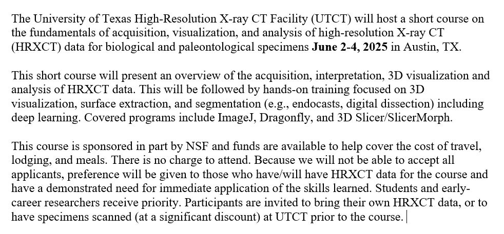
Every year UTCT teaches NSF sponsored short courses on high-resolution X-ray CT, taught by UTCT staff (including me!). Applications for the 2025 courses are now open! See below for the biological/paleontological course info, & we also have a geological one. If you'd like to apply, send me a DM 🩻☢️🦴
April 4, 2025 at 5:52 PM
Every year UTCT teaches NSF sponsored short courses on high-resolution X-ray CT, taught by UTCT staff (including me!). Applications for the 2025 courses are now open! See below for the biological/paleontological course info, & we also have a geological one. If you'd like to apply, send me a DM 🩻☢️🦴
Happy #FossilFriday
A few weeks ago we opened the first exhibit I helped design based on my work on bone histology and taphonomy of a #dicynodont bonebed from Tanzania!
If you are passing by the #FieldMuseum, go check out the #GraingerScienceHub
Huge thanks to @markwitton.bsky.social for 🎨🖼️
A few weeks ago we opened the first exhibit I helped design based on my work on bone histology and taphonomy of a #dicynodont bonebed from Tanzania!
If you are passing by the #FieldMuseum, go check out the #GraingerScienceHub
Huge thanks to @markwitton.bsky.social for 🎨🖼️


March 28, 2025 at 4:04 PM
Happy #FossilFriday
A few weeks ago we opened the first exhibit I helped design based on my work on bone histology and taphonomy of a #dicynodont bonebed from Tanzania!
If you are passing by the #FieldMuseum, go check out the #GraingerScienceHub
Huge thanks to @markwitton.bsky.social for 🎨🖼️
A few weeks ago we opened the first exhibit I helped design based on my work on bone histology and taphonomy of a #dicynodont bonebed from Tanzania!
If you are passing by the #FieldMuseum, go check out the #GraingerScienceHub
Huge thanks to @markwitton.bsky.social for 🎨🖼️
New pub alert! 🚨 and the final installment of papers from my dissertation!
Really happy to share this work on Triassic cynodonts that reveals surprising growth differences, helping to reshape our understanding of stem mammal evolution #ThinSectionThursday #Paleontology #Cynodonts #Triassic
Really happy to share this work on Triassic cynodonts that reveals surprising growth differences, helping to reshape our understanding of stem mammal evolution #ThinSectionThursday #Paleontology #Cynodonts #Triassic
Disparate life histories in coeval Triassic cynodonts and their implications for the evolution of mammalian life histories | Paleobiology | Cambridge Core
Disparate life histories in coeval Triassic cynodonts and their implications for the evolution of mammalian life histories
www.cambridge.org
March 27, 2025 at 1:46 PM
New pub alert! 🚨 and the final installment of papers from my dissertation!
Really happy to share this work on Triassic cynodonts that reveals surprising growth differences, helping to reshape our understanding of stem mammal evolution #ThinSectionThursday #Paleontology #Cynodonts #Triassic
Really happy to share this work on Triassic cynodonts that reveals surprising growth differences, helping to reshape our understanding of stem mammal evolution #ThinSectionThursday #Paleontology #Cynodonts #Triassic
Why do bone histologists use the middle of bones when examining growth patterns? Because mid-shaft record the longest history of growth, seen in this tiny femur where the joint ends are made of spongy bone and only the middle shows growth marks.

February 7, 2025 at 9:17 PM
Why do bone histologists use the middle of bones when examining growth patterns? Because mid-shaft record the longest history of growth, seen in this tiny femur where the joint ends are made of spongy bone and only the middle shows growth marks.
Reposted by Zoe Kulik
If you don't know about the @burkemuseum.bsky.social DIG Field School, it's a great time to learn about this PD program for K-12 educators and classrooms www.burkemuseum.org/education/ed...
Sign up for a Microfossil Workshop by Jan 3! and the DIG Field School applications open soon! #paleo #K12STEM
Sign up for a Microfossil Workshop by Jan 3! and the DIG Field School applications open soon! #paleo #K12STEM
DIG Field School
The DIG Field School connects K-12 STEM teachers with scientific research and researchers through ongoing professional development and teaching curricula.
www.burkemuseum.org
January 2, 2025 at 5:29 PM
If you don't know about the @burkemuseum.bsky.social DIG Field School, it's a great time to learn about this PD program for K-12 educators and classrooms www.burkemuseum.org/education/ed...
Sign up for a Microfossil Workshop by Jan 3! and the DIG Field School applications open soon! #paleo #K12STEM
Sign up for a Microfossil Workshop by Jan 3! and the DIG Field School applications open soon! #paleo #K12STEM
Reposted by Zoe Kulik
New paper alert! This was a fun study examining intraskeletal growth patterns in leopard geckos using fluorescent labeling. Labels administered in ovo are still present up to 4 years post-hatching! doi.org/10.1111/joa....

Bone labeling experiments and intraskeletal growth patterns in captive leopard geckos (Eublepharis macularius)
In this study, we used fluorochrome labels in captive leopard geckos (Eublepharis macularius) to track bone growth and intraskeletal variability from embryonic to adult growth stages. Overall, the ti...
doi.org
November 27, 2024 at 4:26 PM
New paper alert! This was a fun study examining intraskeletal growth patterns in leopard geckos using fluorescent labeling. Labels administered in ovo are still present up to 4 years post-hatching! doi.org/10.1111/joa....
Reposted by Zoe Kulik
Our 2025 #REU applications are now open! Come learn with us next summer at the #AMNH! More information can be found www.amnh.org/research/ric...

Biological Sciences: Research Experience for Undergraduates | AMNH
Approximately eight students gather each summer at the Museum for a ten-week experience working with curators, faculty, and post-doctoral fellows.
www.amnh.org
December 9, 2024 at 8:57 PM
Our 2025 #REU applications are now open! Come learn with us next summer at the #AMNH! More information can be found www.amnh.org/research/ric...
Reposted by Zoe Kulik

🚨New Diamond 💎 Open Access Journal in Paleontology 🦴🐚!🚨
Send us your manuscripts 📜 and publish fully open access 🔓 entirely for free!
#openacess #paleontology #free #palaeontology #diamond #open
Send us your manuscripts 📜 and publish fully open access 🔓 entirely for free!
#openacess #paleontology #free #palaeontology #diamond #open

Hello 👋We are Open Palaeontology, or OPal, a brand new Diamond Open Access journal for palaeontology research.
Our mission is to provide a flexible platform for research that is free to authors and readers.
Read more in our editorial www.openpalaeo.org/article/view... or at www.openpalaeo.org!
Our mission is to provide a flexible platform for research that is free to authors and readers.
Read more in our editorial www.openpalaeo.org/article/view... or at www.openpalaeo.org!
View of Open Palaeontology: a new model of diamond open access journal for palaeontology
www.openpalaeo.org
December 6, 2024 at 9:42 PM
🚨New Diamond 💎 Open Access Journal in Paleontology 🦴🐚!🚨
Send us your manuscripts 📜 and publish fully open access 🔓 entirely for free!
#openacess #paleontology #free #palaeontology #diamond #open
Send us your manuscripts 📜 and publish fully open access 🔓 entirely for free!
#openacess #paleontology #free #palaeontology #diamond #open
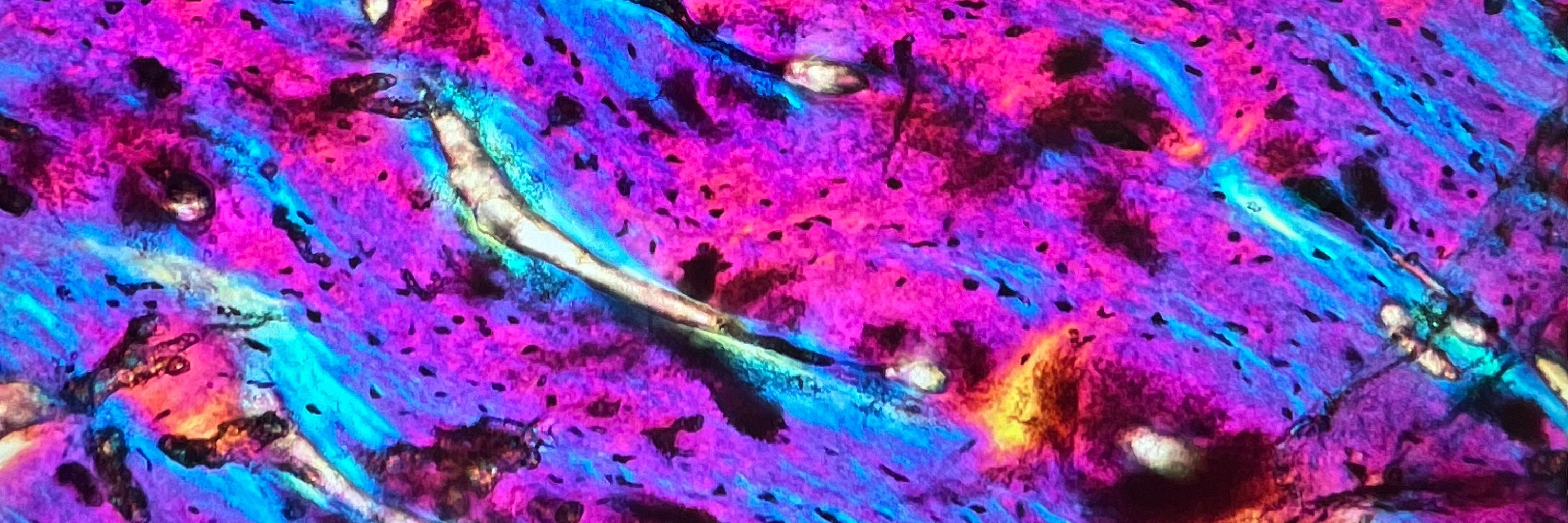
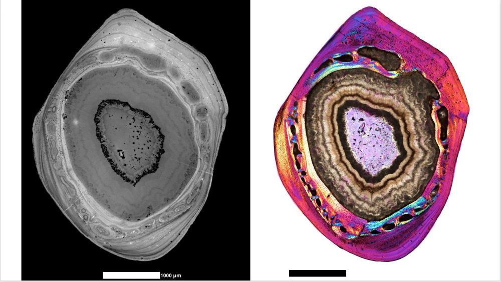
Happy #ThinSectionThursday
Here is a side by side comparison of virtual histology from synchrotron tomography on the left and “traditional” histology from optical microscopy on the right. It’s sometimes hard to tell what’s a growth mark in virtual histology but with this, we can can train our 👀
Here is a side by side comparison of virtual histology from synchrotron tomography on the left and “traditional” histology from optical microscopy on the right. It’s sometimes hard to tell what’s a growth mark in virtual histology but with this, we can can train our 👀
December 5, 2024 at 3:37 PM
Happy #ThinSectionThursday
Here is a side by side comparison of virtual histology from synchrotron tomography on the left and “traditional” histology from optical microscopy on the right. It’s sometimes hard to tell what’s a growth mark in virtual histology but with this, we can can train our 👀
Here is a side by side comparison of virtual histology from synchrotron tomography on the left and “traditional” histology from optical microscopy on the right. It’s sometimes hard to tell what’s a growth mark in virtual histology but with this, we can can train our 👀
Reposted by Zoe Kulik
Job alert! Two Full-time Assistant Teaching Professor positions in our Department (UW Biology). Come join us! Please share: apply.interfolio.com/147513
November 22, 2024 at 4:18 PM
Job alert! Two Full-time Assistant Teaching Professor positions in our Department (UW Biology). Come join us! Please share: apply.interfolio.com/147513
A late night #ThinSectionThursday
Resisting that “we’ll pick it up in the new year” mentality with cool collaborations that have data that looks this good
Resisting that “we’ll pick it up in the new year” mentality with cool collaborations that have data that looks this good

November 22, 2024 at 3:59 AM
A late night #ThinSectionThursday
Resisting that “we’ll pick it up in the new year” mentality with cool collaborations that have data that looks this good
Resisting that “we’ll pick it up in the new year” mentality with cool collaborations that have data that looks this good
Paleobluesky has risen!
So happy to be here and share a sneak peak of some micro-CT scans. Check out the 3D histology of some truly tiny Mesozoic mammal fossils 🤩 🐭🐹🐿️
#fossilfriday #fossil #zeissxradia #amnh #watchthisspace
So happy to be here and share a sneak peak of some micro-CT scans. Check out the 3D histology of some truly tiny Mesozoic mammal fossils 🤩 🐭🐹🐿️
#fossilfriday #fossil #zeissxradia #amnh #watchthisspace


November 15, 2024 at 11:26 PM
Paleobluesky has risen!
So happy to be here and share a sneak peak of some micro-CT scans. Check out the 3D histology of some truly tiny Mesozoic mammal fossils 🤩 🐭🐹🐿️
#fossilfriday #fossil #zeissxradia #amnh #watchthisspace
So happy to be here and share a sneak peak of some micro-CT scans. Check out the 3D histology of some truly tiny Mesozoic mammal fossils 🤩 🐭🐹🐿️
#fossilfriday #fossil #zeissxradia #amnh #watchthisspace
Reposted by Zoe Kulik
Come work with us! We are hiring a new #curator in invertebrate paleontology! careers.amnh.org/postings/4301 #academicjobs #tenuretrack #AMNH
Assistant Curator/Professor of Invertebrate Paleontology
The Division of Paleontology at the American Museum of Natural History (AMNH) seeks an Assistant Curator of Invertebrate Paleontology to start on or after July 1, 2025.The successful candidate will be...
careers.amnh.org
November 14, 2024 at 7:56 PM
Come work with us! We are hiring a new #curator in invertebrate paleontology! careers.amnh.org/postings/4301 #academicjobs #tenuretrack #AMNH
#ThinSectionThursday brought to you by this stunning watercolor painting by Leonie Schön. Check out her histology artwork!
lamellipodiumart.com
lamellipodiumart.com

November 14, 2024 at 5:56 PM
#ThinSectionThursday brought to you by this stunning watercolor painting by Leonie Schön. Check out her histology artwork!
lamellipodiumart.com
lamellipodiumart.com
I’m new to this online paleo community and am looking forward to getting connected. If you’re going to #2024SVP next week, stop by my talk on Wed. at 8am in the mammalian life history evolution symposium for a deep dive into 3D bone histology from some of the earliest non-mammalian synapsids!

October 25, 2024 at 2:29 AM
I’m new to this online paleo community and am looking forward to getting connected. If you’re going to #2024SVP next week, stop by my talk on Wed. at 8am in the mammalian life history evolution symposium for a deep dive into 3D bone histology from some of the earliest non-mammalian synapsids!