


Structural Biology Communications
@actacrystf.iucr.org
Acta Crystallographica Section F: Structural Biology Communications is a structural biology communications journal published by the IUCr. http://journals.iucr.org/f
First issue of 2026 is out: journals.iucr.org/f/issues/202...
On the cover: single surface substitutions improve the crystallizability and diffraction properties of a flexible two-domain protein, even if they increase the entropy of the respective residue. Read more at
doi.org/10.1107/S205...
On the cover: single surface substitutions improve the crystallizability and diffraction properties of a flexible two-domain protein, even if they increase the entropy of the respective residue. Read more at
doi.org/10.1107/S205...
January 5, 2026 at 1:40 PM
First issue of 2026 is out: journals.iucr.org/f/issues/202...
On the cover: single surface substitutions improve the crystallizability and diffraction properties of a flexible two-domain protein, even if they increase the entropy of the respective residue. Read more at
doi.org/10.1107/S205...
On the cover: single surface substitutions improve the crystallizability and diffraction properties of a flexible two-domain protein, even if they increase the entropy of the respective residue. Read more at
doi.org/10.1107/S205...
CryoSift is a platform-independent convolutional neural network tool for assessing the quality of 2D averages to enable the automatic selection of suitable particles for high-resolution reconstructions #Automation #DataProcessing #CryoEM doi.org/10.1107/S205...

December 18, 2025 at 3:32 PM
CryoSift is a platform-independent convolutional neural network tool for assessing the quality of 2D averages to enable the automatic selection of suitable particles for high-resolution reconstructions #Automation #DataProcessing #CryoEM doi.org/10.1107/S205...
The rigid single-domain structure and reproducible crystallization behavior of PF1765 from P. furiosus indicate potential use as a fusion domain to aid the crystallization of membrane proteins or complexes #PF1765 #UPF0235 #ProteinCrystallography doi.org/10.1107/S205...

December 17, 2025 at 2:01 PM
The rigid single-domain structure and reproducible crystallization behavior of PF1765 from P. furiosus indicate potential use as a fusion domain to aid the crystallization of membrane proteins or complexes #PF1765 #UPF0235 #ProteinCrystallography doi.org/10.1107/S205...
Structural analysis of apo and UDP-bound tetrahydrobiopterin glucosyltransferase revealed features at the active-site pocket differing from other retaining GT-B glycosyltransferases #Tetrahydrobiopterin #GTBGlycosyltransferase #Crystallography doi.org/10.1107/S205...

December 16, 2025 at 2:06 PM
Structural analysis of apo and UDP-bound tetrahydrobiopterin glucosyltransferase revealed features at the active-site pocket differing from other retaining GT-B glycosyltransferases #Tetrahydrobiopterin #GTBGlycosyltransferase #Crystallography doi.org/10.1107/S205...
In the coenzyme-bound structure of short-chain dehydrogenase from Brucella ovis, two different orientations of the NAD+ coenzyme are fortuitously observed #ShortChainDehydrogenaseReductase #ResearchExperienceForUndergraduates doi.org/10.1107/S205...

December 15, 2025 at 3:00 PM
In the coenzyme-bound structure of short-chain dehydrogenase from Brucella ovis, two different orientations of the NAD+ coenzyme are fortuitously observed #ShortChainDehydrogenaseReductase #ResearchExperienceForUndergraduates doi.org/10.1107/S205...
Our last issue of 2026 is available: journals.iucr.org/f/issues/202...
On the cover: two different orientations of the NAD coenzyme are fortuitously observed in the NAD+-bound structure of a short-chain dehydrogenase from Brucella ovis. Find out more at doi.org/10.1107/S205...
On the cover: two different orientations of the NAD coenzyme are fortuitously observed in the NAD+-bound structure of a short-chain dehydrogenase from Brucella ovis. Find out more at doi.org/10.1107/S205...
December 3, 2025 at 1:31 PM
Our last issue of 2026 is available: journals.iucr.org/f/issues/202...
On the cover: two different orientations of the NAD coenzyme are fortuitously observed in the NAD+-bound structure of a short-chain dehydrogenase from Brucella ovis. Find out more at doi.org/10.1107/S205...
On the cover: two different orientations of the NAD coenzyme are fortuitously observed in the NAD+-bound structure of a short-chain dehydrogenase from Brucella ovis. Find out more at doi.org/10.1107/S205...
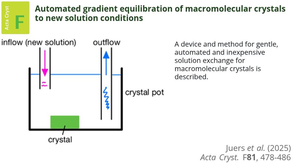
A gentle, automated, inexpensive and open-source device and method for changing the ambient solution of a macromolecular crystal #Crystal #Solution #Cracking doi.org/10.1107/S205...
November 13, 2025 at 3:29 PM
A gentle, automated, inexpensive and open-source device and method for changing the ambient solution of a macromolecular crystal #Crystal #Solution #Cracking doi.org/10.1107/S205...
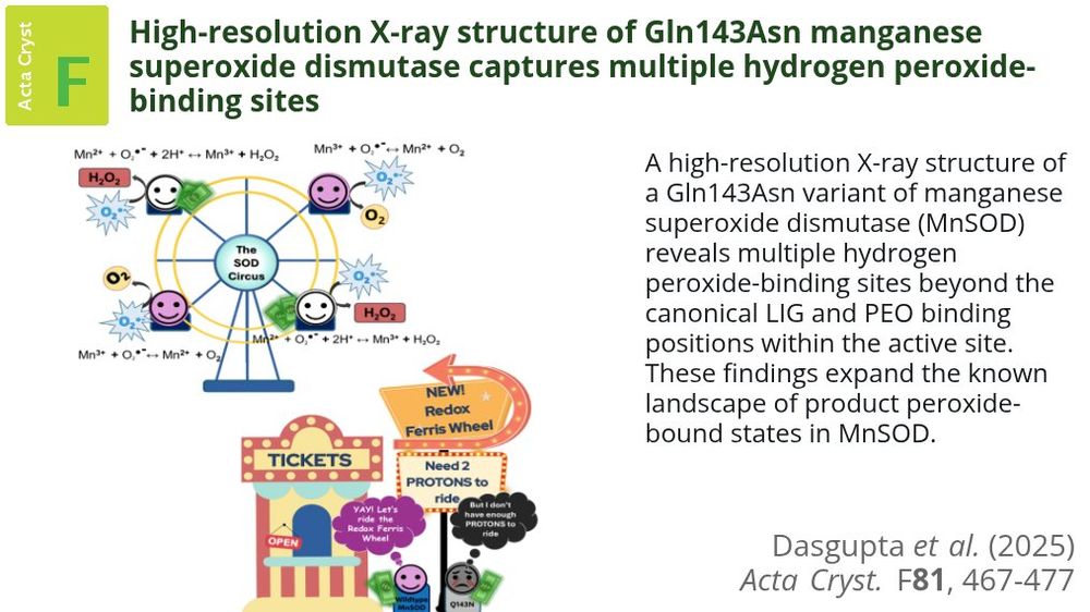
A kinetically impaired Gln143Asn MnSOD variant is used to trap and explore additional hydrogen peroxide-binding sites beyond the second-shell solvent gate #ManganeseSuperoxideDismutases #PeroxideBinding #Metalloenzymes doi.org/10.1107/S205...
November 12, 2025 at 4:06 PM
A kinetically impaired Gln143Asn MnSOD variant is used to trap and explore additional hydrogen peroxide-binding sites beyond the second-shell solvent gate #ManganeseSuperoxideDismutases #PeroxideBinding #Metalloenzymes doi.org/10.1107/S205...
The first crystal structure of FucWf4, a newly characterized fucosidase that has structural similarity to GH29 enzymes but is currently unclassified, is presented #GlycosideHydrolases #Fucoidan #Fucosidase doi.org/10.1107/S205...

November 11, 2025 at 1:45 PM
The first crystal structure of FucWf4, a newly characterized fucosidase that has structural similarity to GH29 enzymes but is currently unclassified, is presented #GlycosideHydrolases #Fucoidan #Fucosidase doi.org/10.1107/S205...
Structural investigations of unliganded murine KHK-A revealed the adoption of two conformations similar to those adopted by the human ortholog, suggesting this structural feature is conserved across species #PfkBFamily #Moonlighting #ProteinKinases doi.org/10.1107/S205...

November 10, 2025 at 2:16 PM
Structural investigations of unliganded murine KHK-A revealed the adoption of two conformations similar to those adopted by the human ortholog, suggesting this structural feature is conserved across species #PfkBFamily #Moonlighting #ProteinKinases doi.org/10.1107/S205...
November issue out now: journals.iucr.org/f/issues/202...
On the cover: the structure of the kinetically slow and catalytically impaired Gln143Asn mutant of human mitochondrial MnSOD leads to the observation of transient hydrogen peroxide-bound states. More at shorturl.at/41Mb9
On the cover: the structure of the kinetically slow and catalytically impaired Gln143Asn mutant of human mitochondrial MnSOD leads to the observation of transient hydrogen peroxide-bound states. More at shorturl.at/41Mb9

November 5, 2025 at 1:25 PM
November issue out now: journals.iucr.org/f/issues/202...
On the cover: the structure of the kinetically slow and catalytically impaired Gln143Asn mutant of human mitochondrial MnSOD leads to the observation of transient hydrogen peroxide-bound states. More at shorturl.at/41Mb9
On the cover: the structure of the kinetically slow and catalytically impaired Gln143Asn mutant of human mitochondrial MnSOD leads to the observation of transient hydrogen peroxide-bound states. More at shorturl.at/41Mb9
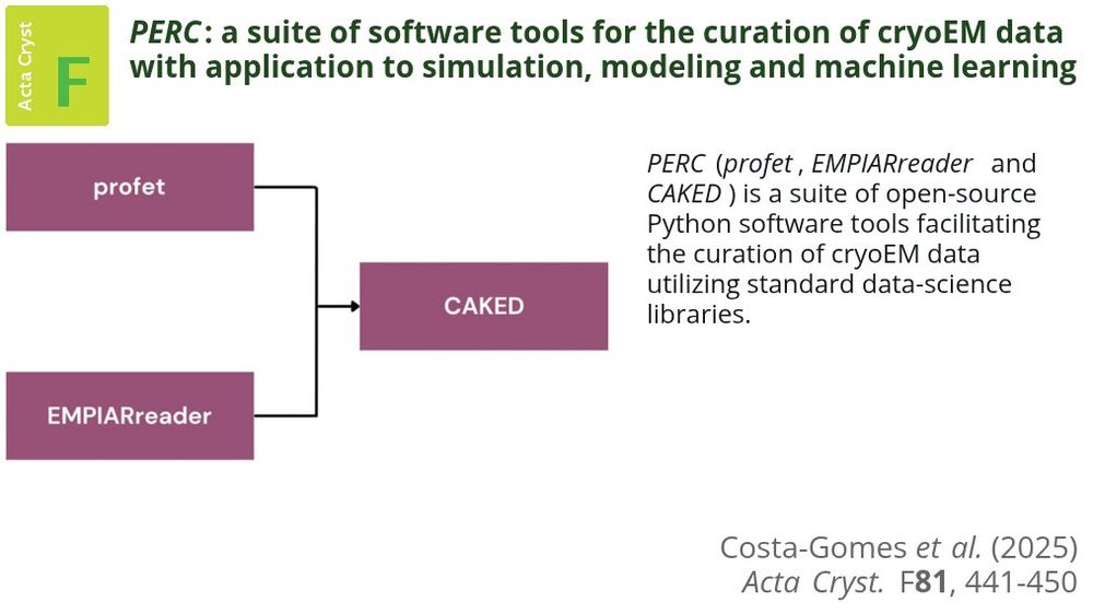
Tools are presented that are useful for collating existing public cryoEM data sets and/or creating new synthetic cryoEM data sets to aid the development of novel data processing and interpretation algorithms #CryoEM #ElectronCryomicroscopy #Python doi.org/10.1107/S205...
October 22, 2025 at 2:16 PM
Tools are presented that are useful for collating existing public cryoEM data sets and/or creating new synthetic cryoEM data sets to aid the development of novel data processing and interpretation algorithms #CryoEM #ElectronCryomicroscopy #Python doi.org/10.1107/S205...
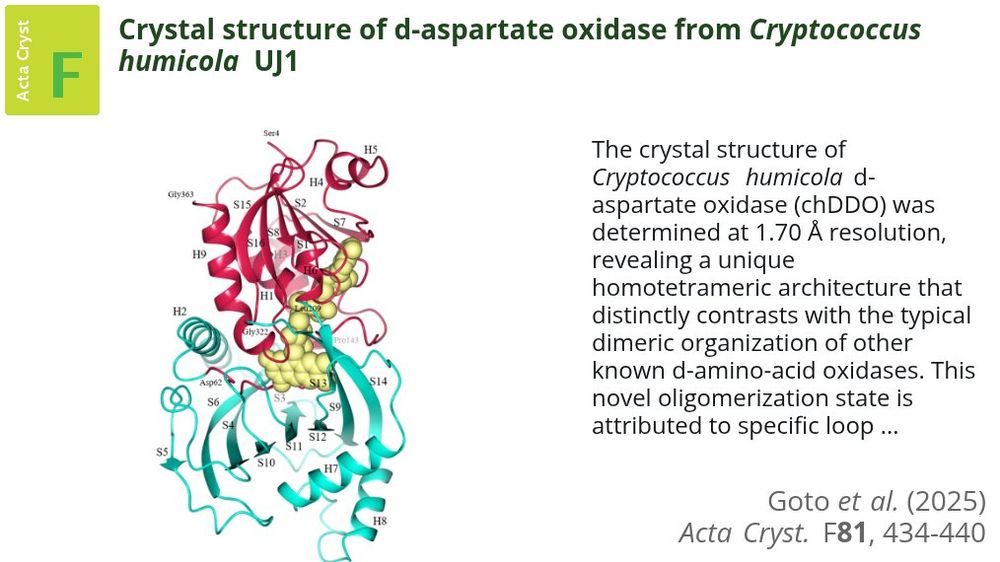
D-Aspartate oxidase from Cryptococcus humicola UJ1 reveals a unique homotetrameric organization due to distinct interfacial loop arrangements #DAspartateOxidase #CrystalStructure #FADDependentEnzymes doi.org/10.1107/S205...
October 21, 2025 at 3:00 PM
D-Aspartate oxidase from Cryptococcus humicola UJ1 reveals a unique homotetrameric organization due to distinct interfacial loop arrangements #DAspartateOxidase #CrystalStructure #FADDependentEnzymes doi.org/10.1107/S205...
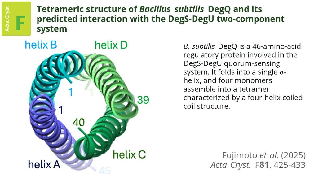
The structure of B. subtilis DegQ from the DegS–DegU two-component system provide structural insight into DegQ oligomerization and its potential role in modulating DegS autophosphorylation and DegU binding #BacillusSubtilis #CrystalStructure #DegQ doi.org/10.1107/S205...
October 20, 2025 at 12:52 PM
The structure of B. subtilis DegQ from the DegS–DegU two-component system provide structural insight into DegQ oligomerization and its potential role in modulating DegS autophosphorylation and DegU binding #BacillusSubtilis #CrystalStructure #DegQ doi.org/10.1107/S205...
Four cyanobacterial glycosyl transferase family 35 α-glucan phosphorylases of types I and II were biochemically characterized and one was crystallized and crystallographically analyzed #Cyanobacteria #CyanobacterialStarch #Glycogen doi.org/10.1107/S205...

October 9, 2025 at 3:04 PM
Four cyanobacterial glycosyl transferase family 35 α-glucan phosphorylases of types I and II were biochemically characterized and one was crystallized and crystallographically analyzed #Cyanobacteria #CyanobacterialStarch #Glycogen doi.org/10.1107/S205...
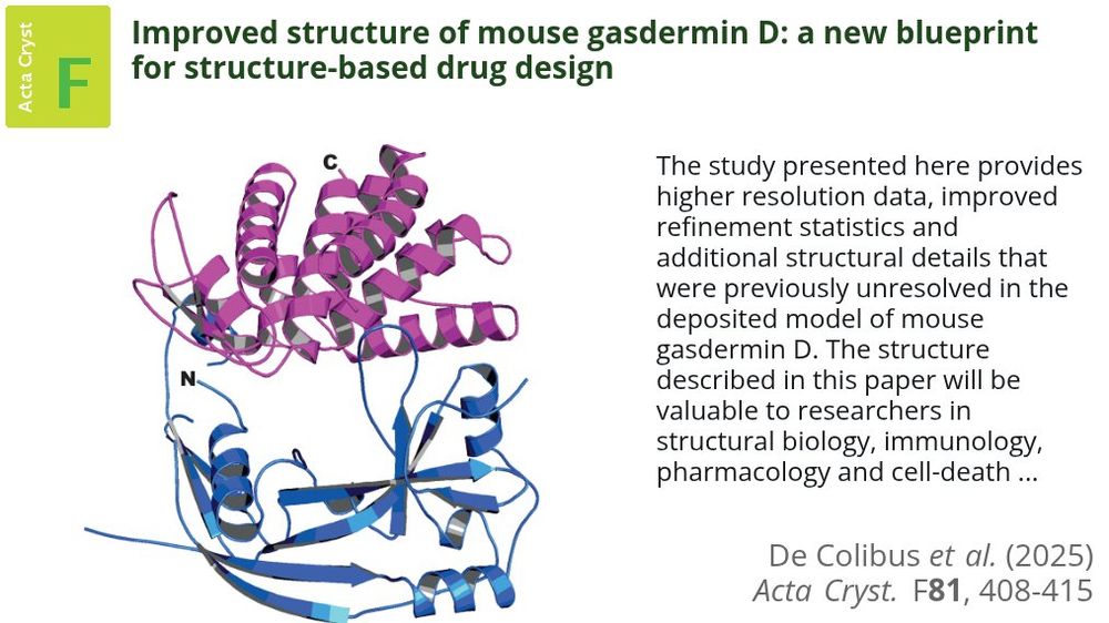
The higher resolution, significantly improved apo crystal structure of mouse gasdermin D will be beneficial for structure-based drug-design approaches towards this important pharmacological target #Pyroptosis #Inflammation #GasderminD doi.org/10.1107/S205...
October 8, 2025 at 1:35 PM
The higher resolution, significantly improved apo crystal structure of mouse gasdermin D will be beneficial for structure-based drug-design approaches towards this important pharmacological target #Pyroptosis #Inflammation #GasderminD doi.org/10.1107/S205...
The SAMPREP workshop at the 2023 ACA Annual Meeting in Baltimore was designed to address the need to consoiidate cross-disciplinary best practices for sample preparation into a single forum #SamplePreparation #StructuralBiology #CryoEM doi.org/10.1107/S205...

October 7, 2025 at 12:48 PM
The SAMPREP workshop at the 2023 ACA Annual Meeting in Baltimore was designed to address the need to consoiidate cross-disciplinary best practices for sample preparation into a single forum #SamplePreparation #StructuralBiology #CryoEM doi.org/10.1107/S205...
Take a look at our October issue: journals.iucr.org/f/issues/202...
The cover shows the crystal structure of Cryptococcus humicola D-aspartate oxidase, which has a unique homotetrameric architecture in contrast to other known D-amino-acid oxidases. Read more at doi.org/10.1107/S205...
The cover shows the crystal structure of Cryptococcus humicola D-aspartate oxidase, which has a unique homotetrameric architecture in contrast to other known D-amino-acid oxidases. Read more at doi.org/10.1107/S205...
October 2, 2025 at 1:45 PM
Take a look at our October issue: journals.iucr.org/f/issues/202...
The cover shows the crystal structure of Cryptococcus humicola D-aspartate oxidase, which has a unique homotetrameric architecture in contrast to other known D-amino-acid oxidases. Read more at doi.org/10.1107/S205...
The cover shows the crystal structure of Cryptococcus humicola D-aspartate oxidase, which has a unique homotetrameric architecture in contrast to other known D-amino-acid oxidases. Read more at doi.org/10.1107/S205...
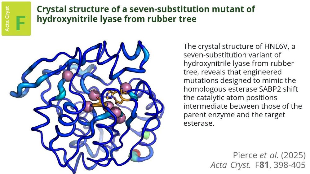
The structure of a 7-substitution mutant of hydroxynitrile lyase shows that targeted amino-acid substitutions can shift catalytic geometries towards those of evolutionarily related enzymes #EngineeredProteins #HydroxynitrileLyases #HydrolaseFold doi.org/10.1107/S205...
September 15, 2025 at 3:14 PM
The structure of a 7-substitution mutant of hydroxynitrile lyase shows that targeted amino-acid substitutions can shift catalytic geometries towards those of evolutionarily related enzymes #EngineeredProteins #HydroxynitrileLyases #HydrolaseFold doi.org/10.1107/S205...
The World Directory of Crystallographers (WDC) offers an unparalleled compilation of expertise in the field.
It’s free to join and open to all crystallographers and other scientists employing crystallographic methods.
Add your name and be part of the mosaic:
🔗 www.iucr.org/people/wdc
It’s free to join and open to all crystallographers and other scientists employing crystallographic methods.
Add your name and be part of the mosaic:
🔗 www.iucr.org/people/wdc

September 12, 2025 at 1:05 PM
The World Directory of Crystallographers (WDC) offers an unparalleled compilation of expertise in the field.
It’s free to join and open to all crystallographers and other scientists employing crystallographic methods.
Add your name and be part of the mosaic:
🔗 www.iucr.org/people/wdc
It’s free to join and open to all crystallographers and other scientists employing crystallographic methods.
Add your name and be part of the mosaic:
🔗 www.iucr.org/people/wdc
Crystal structures of a clinically used histone deacetylase inhibitor, vorinostat, bound to the previously unidentified off-target proteins carbonic anhydrases II and IX are reported #HDACInhibitors #SAHA #Vorinostat doi.org/10.1107/S205...

September 11, 2025 at 1:31 PM
Crystal structures of a clinically used histone deacetylase inhibitor, vorinostat, bound to the previously unidentified off-target proteins carbonic anhydrases II and IX are reported #HDACInhibitors #SAHA #Vorinostat doi.org/10.1107/S205...
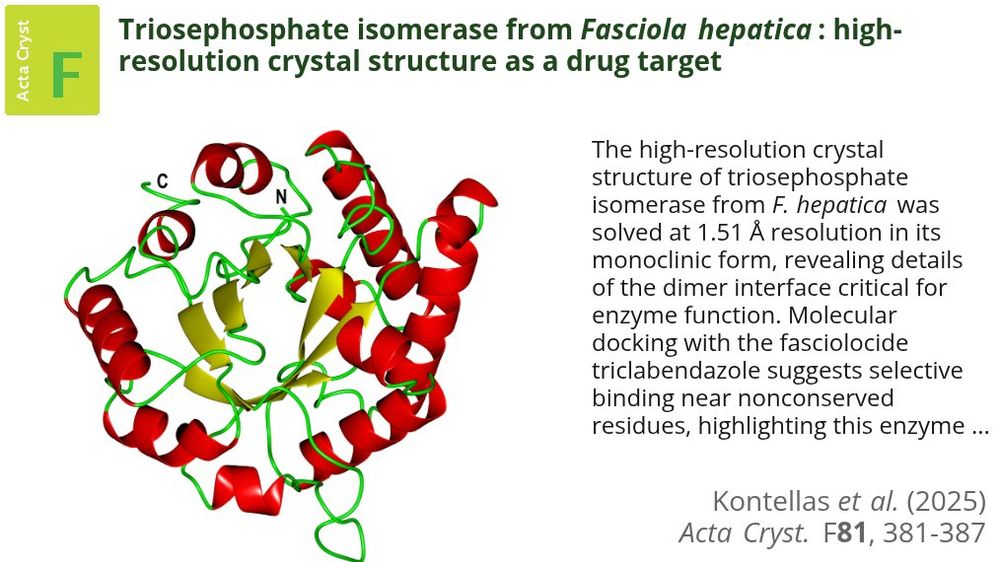
The crystal structure of Fasciola hepatica TPI has been solved at 1.51 Å and used to perform molecular-docking studies with the most successful fasciolocide triclabendazole #FasciolaHepatica #TriosephosphateIsomerase #TPI doi.org/10.1107/S205...
September 10, 2025 at 2:37 PM
The crystal structure of Fasciola hepatica TPI has been solved at 1.51 Å and used to perform molecular-docking studies with the most successful fasciolocide triclabendazole #FasciolaHepatica #TriosephosphateIsomerase #TPI doi.org/10.1107/S205...
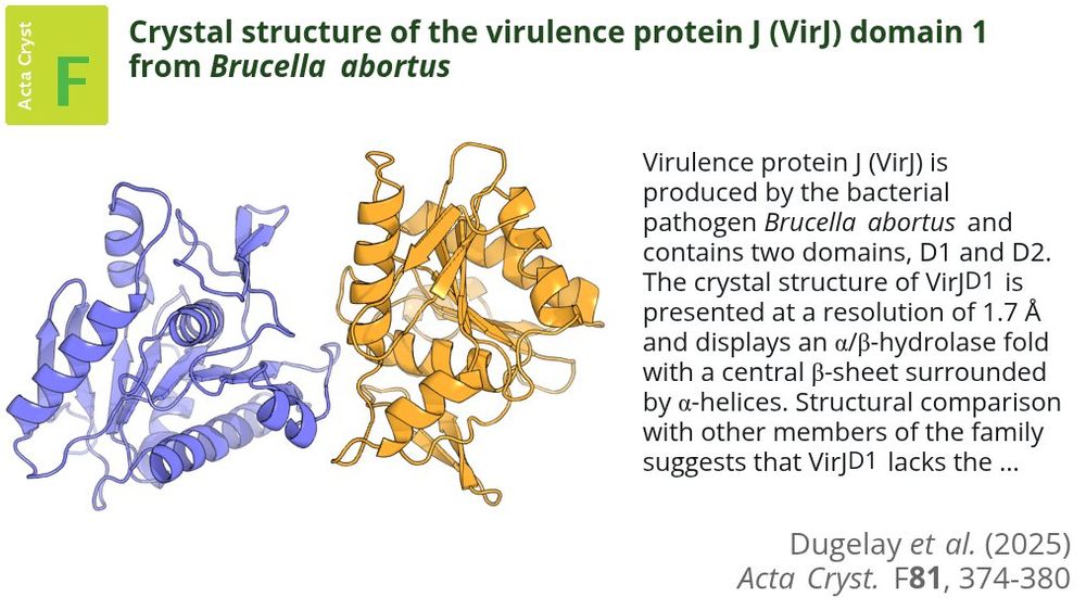
Virulence protein J domain 1 maintains the canonical α/β-hydrolase fold but is likely to lack a functional catalytic triad, suggesting functional divergence from classical hydrolases of this superfamily #BacterialPathogens #TypeIVSecretionSystem doi.org/10.1107/S205...
September 9, 2025 at 2:50 PM
Virulence protein J domain 1 maintains the canonical α/β-hydrolase fold but is likely to lack a functional catalytic triad, suggesting functional divergence from classical hydrolases of this superfamily #BacterialPathogens #TypeIVSecretionSystem doi.org/10.1107/S205...
The first high-resolution structure of the folded domains of Xrs2 from S. cerevisiae will advance understanding of its functions in the DNA damage response and telomere-length maintenance @UMNews #Nbs1Xrs2 #DNADoubleStrandBreaks #DNADamageRepair doi.org/10.1107/S205...

September 8, 2025 at 1:28 PM
The first high-resolution structure of the folded domains of Xrs2 from S. cerevisiae will advance understanding of its functions in the DNA damage response and telomere-length maintenance @UMNews #Nbs1Xrs2 #DNADoubleStrandBreaks #DNADamageRepair doi.org/10.1107/S205...
Our September issue is available! tinyurl.com/58x22mpx
The cover shows the crystal structure of the N-terminal domain 1 of virulence protein J from Brucella abortus, which adopts an α/β-hydrolase fold but lacks the characteristic catalytic triad: see tinyurl.com/bdduy23d
The cover shows the crystal structure of the N-terminal domain 1 of virulence protein J from Brucella abortus, which adopts an α/β-hydrolase fold but lacks the characteristic catalytic triad: see tinyurl.com/bdduy23d
September 3, 2025 at 2:20 PM
Our September issue is available! tinyurl.com/58x22mpx
The cover shows the crystal structure of the N-terminal domain 1 of virulence protein J from Brucella abortus, which adopts an α/β-hydrolase fold but lacks the characteristic catalytic triad: see tinyurl.com/bdduy23d
The cover shows the crystal structure of the N-terminal domain 1 of virulence protein J from Brucella abortus, which adopts an α/β-hydrolase fold but lacks the characteristic catalytic triad: see tinyurl.com/bdduy23d