Ana Martínez Gómez
@brainbyana.bsky.social
24 followers 71 following 54 posts
PhD student Brain & Neurogenesis | Evolution of the Brain | Genomics
Posts Media Videos Starter Packs
Pinned
brainbyana.bsky.social
Here’s the first paper I contributed to: “Expression of SATB1 and SATB2 in the brain of bony fishes: what fish reveal about evolution”
DOI: doi.org/10.1007/s004...
Reposted by Ana Martínez Gómez
loretamedinah.bsky.social
Happy to share our new article with Júlia Freixes, Fatma Abdel-Rahman, Roberto Nebbia and @edesfilis.bsky.social on postnatal plasticity in olfactory areas of the juvenile swine, published in Brain, Structure and Function link.springer.com/article/10.1...
Postnatal plasticity in the olfactory system of the juvenile swine brain - Brain Structure and Function
Swine have an excellent sense of smell and highly complex olfactory brain structures, which play a crucial role in their complex social interactions. In other mammals the olfactory system is known to exhibit significant plasticity, even during adulthood. The aim of this study was to investigate postnatal plasticity in olfactory areas of juvenile swine brains by studying immature cells immunoreactive for the microtubule-associated protein doublecortin (DCX). Using immunofluorescence, we studied DCX coexpression with the cell proliferation marker Ki-67, and different neuronal markers. Our results show the existence of numerous DCX + cells throughout the olfactory pallial areas. In some of them, we found DCX+/Ki-67 + coexpressing cells, suggesting that they were proliferating. Some of these proliferating cells were grouped in tangentially-oriented migratory-like chains, forming the rostral migratory stream to anterior olfactory area and olfactory bulb. Moreover, chains of DCX + cells were found in the external capsule and white matter adjacent to the temporal horn of the ventricle. Chains of DCX + cells were observed crossing the internal layers of the piriform and entorhinal cortices. In layer II of these cortices, DCX + cells of varying maturity degrees and neuronal phenotypes (including NeuN expression) were present. This suggests the existence of multiple migratory streams along the anteroposterior axis. Most DCX + immature cells in the migratory chains and in the anterior olfactory area, piriform and entorhinal cortices expressed the transcription factor Brn2 (Pou3f2), suggesting the incorporation of new glutamatergic neurons in these areas. Together, these results highlight the interest of swine to study the role of postnatal brain plasticity and their potential for regeneration in large, gyrencephalic brains.
link.springer.com
brainbyana.bsky.social
13. Mahmoudi & Cairns 2019, Sci Rep, doi.org/10.1038/s415...
14. Westholm et al. 2014, Cell Rep, DOI: 10.1016/j.celrep.2014.10.062
15. Gruner et al. 2016, Sci Rep, doi.org/10.1038/srep...
brainbyana.bsky.social
10. Chen & Sarnow 1995, Science, DOI: 10.1126/science.7536344
11. You et al. 2015, Nat Neurosci, doi.org/10.1038/nn.3...
12. Rybak-Wolf et al. 2014, Mol Cell, DOI: 10.1016/j.molcel.2015.03.027
brainbyana.bsky.social
5. Hansen et al. 2013, Nature,https://doi.org/10.1038/nature11993
6. Hollensen et al. 2020, Elife, doi.org/10.7554/eLif...
7. Chen et al. 2018, Genome Biol, doi.org/10.1186/s130...
8. Tay & Pek 2017, Curr Biol, DOI: 10.1016/j.cub.2017.02.040
9. Abe et al. 2015, Sci Rep, doi.org/10.1038/srep...
brainbyana.bsky.social
Bibliography:

1. Liu & Chen 2022, Cell, DOI: 10.1016/j.cell.2022.04.021
2. Lasda & Parker 2014, RNA,http://www.rnajournal.org/cgi/doi/10.1261/rna.047126.114
3. Li et al. 2015, Nat Struct Mol Biol, doi.org/10.1038/nsmb...
4. Zhang et al. 2013, Mol Cell, DOI: 10.1016/j.molcel.2013.08.017
brainbyana.bsky.social
📚 For a deeper dive into circRNAs in brain development and disease, check out: “Circular RNAs: Emblematic Players of Neurogenesis and Neurodegeneration”

DOI: doi.org/10.3390/ijms...
brainbyana.bsky.social
🧠 circRNAs are highly enriched in neural tissue (~20% of the transcriptome), localize in cytoplasm and synapses, increase during differentiation and aging, and show spatiotemporal expression independent of linear mRNAs 11-15.
brainbyana.bsky.social
🧩 circRNAs can be translated via internal initiation, challenging their classic label as non-coding 9,10.
This expands their regulatory potential, though the functional roles of endogenously produced proteins remain largely unclear.
brainbyana.bsky.social
circRNAs have diverse roles:
They primarily act as miRNA sponges, regulating gene expression 5.
They also modulate proteins—as decoys, scaffolds, and recruiters—and in the nucleus, boost host gene transcription, collectively influencing cell cycle, apoptosis, and cell survival 1,3,4,6,7,8.
brainbyana.bsky.social
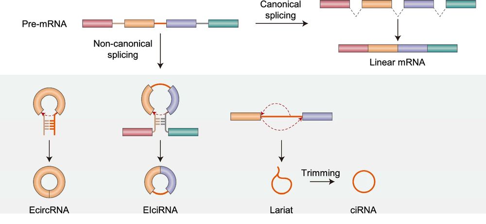
🌀 circRNAs lack a 5′ cap and 3′ poly(A) tail and are generated through a noncanonical splicing process called back-splicing 1,2.
They are classified as ecRNAs, ciRNAs, or EIciRNAs, depending on whether they contain exons, introns, or both 3-4.
brainbyana.bsky.social
🧬 Circular RNAs (circRNAs) are single-stranded molecules once classified as non-coding, now emerging as crucial regulators of gene expression and protein dynamics.
brainbyana.bsky.social
Let's talk about circular RNA!! 🧬🧬

Read the review:
“Molecular mechanisms of circular RNA translation” DOI: doi.org/10.1038/s122...

#MolecularBiology #Genomics #Science #Transcriptomics #RNAbiology #GeneExpression
brainbyana.bsky.social
13. Gabi et al. 2010, Brain Behav Evol, doi:10.1159/000319872
brainbyana.bsky.social
9. Cunha et al. 2022, Front Neuroanat, doi:10.3389/fnana.2022.1048261
10. Teles et al. 2012, J Comp Physiol A, doi:10.1007/s00359-012-0721-6
11. Zupanc 1999, J Exp Biol, doi:10.1242/jeb.202.10.1435
12. Zupanc 2021, J Exp Biol, doi:10.1242/jeb.226357
brainbyana.bsky.social
5. Olkowicz et al. 2016, PNAS, doi:10.1073/pnas.1517131113
6. Kverková et al. 2022, PNAS, doi:10.1073/pnas.2121624119
7. Herculano-Houzel et al. 2011, Brain Behav Evol, doi:10.1159/000330825
8. Herculano-Houzel et al. 2015, Front Neuroanat, doi:10.3389/fnana.2015.00064
brainbyana.bsky.social
Bibliography:

1. Jerison 1973, Evolution of the Brain and Intelligence, Academic Press
2. Marhounová et al. 2019, Evolution, doi:10.1111/evo.13805
3. Herculano-Houzel et al. 2007, PNAS, doi:10.1073/pnas.0611396104
4. Herculano-Houzel et al. 2014, Front Neuroanat, doi:10.3389/fnana.2014.00077
brainbyana.bsky.social
Over generations, step changes → cohorts diverge in brain, body & neuron number.
Some clades (primates, parrots, songbirds) evolve bigger brains with more neurons; others (rodents, sauropsids, mammals) grow brains faster than neurons → lower density 3-6,13.
👉 Plasticity drives brain diversity.
brainbyana.bsky.social
Reaction norm model:
🌍 Environmental step changes (like density) shift whole cohorts into new scaling regimes.
🧬 Within a cohort, individual-level factors (genetics, epigenetics, microenvironment) decouple body, brain, and neuron numbers.
brainbyana.bsky.social
This pattern contradicts the “numerical matching” hypothesis, which predicts body, brain, and neuron number should always scale together 11, 12.
Instead, brain size and neuron number may be regulated by different mechanisms.
brainbyana.bsky.social
Across cohorts (different densities):
🐟 Larger fish → larger brains with up to 20× more neurons.
Neuron density = constant or decreasing.
📐 This mirrors well-known interspecies scaling patterns, even though all fish came from the same stock.
brainbyana.bsky.social
Within a cohort (same density):
🐟 Larger fish → larger brains, but not more neurons.
Animals with more neurons had higher neuron density instead.
⚡ This extends to fish the brain-scaling pattern seen within individuals of the same species in rodents and birds.
brainbyana.bsky.social
Across vertebrates, bigger species → bigger brains with more neurons 1-6.
But within a species, this often breaks down 3,7-9.
Tilapia are ideal to test: they keep growing + have adult neurogenesis 10.
Researchers raised them at low vs. high density → up to 30× body size variation in same-age fish.
Reposted by Ana Martínez Gómez
laurentformery.bsky.social
Sea urchin metamorphosis is maybe one of the most radical event in developmental biology. In just about an hour, these little pluteus larvae completely reorganize their entire body plan.

Very happy that this video got awarded an honorable mention by #NikonSmallWorld 😃