Carolin Klose
@carolinklose.bsky.social
110 followers 160 following 22 posts
PhD student in Munich with Matthias Feige (TUM) and Brenda Schulman (MPI Biochemistry) Proteostasis and membrane protein enthusiast Boehringer Ingelheim Fonds fellow
Posts Media Videos Starter Packs
carolinklose.bsky.social
Thank you! It's EMC time 😉
carolinklose.bsky.social
Thank you to everyone involved in the Schulman @mpibiochem.bsky.social , Feige @tum.de , @fenech-lab.bsky.social , and Schuldiner labs! 🥳
carolinklose.bsky.social
A potential model: the conserved EMC:Spf1 supercomplex spatially couples insertion and extraction. Juxtaposed functional sites form a shared cavity, enabling substrate handover and discrimination. Spf1's nucleotide state may regulate access to this cavity, coordinating insertion and extraction.
carolinklose.bsky.social
Translocation by Spf1 is coupled to ATP hydrolysis. To probe its functional cycle in the EMC:Spf1 complex, we determined structures in the E1-ATP and E1-P states.
ATP binding stabilizes Spf1’s “arm” domain, contacting EMC’s cytoplasmic cap above the insertase cavity, closing the composite cavity.
carolinklose.bsky.social
The high stability of the yeast EMC:Spf1 complex suggests a key functional relationship. Using endogenous tagging, mass spectrometry, modeling and experimental validation, we show that a similar complex exists in human cells between EMC and ATP13A1.
carolinklose.bsky.social
The main site of interaction is confined to a lumenal interface (“lumenal dock”) involving EMC7, EMC10, and the charged lumenal surface of Spf1.
carolinklose.bsky.social
The architecture of this supercomplex reveals juxtaposed functional sites for TMD insertion (EMC) and extraction (Spf1), forming a large composite intramembrane cavity.
carolinklose.bsky.social
We found that at endogenous levels in yeast, the EMC forms a stoichiometric complex with Spf1. Spf1 is a TMD dislocase, the biochemical counterpart to EMC's role as insertase. To gain more insights into this intriguing supercomplex, we determined the EMC:Spf1 structure by cryo-EM.
carolinklose.bsky.social
Super excited to share our new #preprint on #BioRxiv
We reveal the structural basis of a partnership between the ER membrane complex (EMC) and the P5A-ATPase Spf1 — an insertase–dislocase duo that coordinates membrane protein biogenesis and quality control.
www.biorxiv.org/content/10.1...
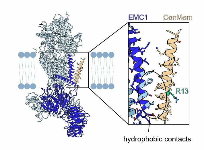
Structural basis of an EMC:Spf1 insertase-dislocase complex in the eukaryotic endoplasmic reticulum
Most eukaryotic membrane proteins are inserted into the membrane at the endoplasmic reticulum (ER). This essential but error-prone process relies on molecular quality control machineries to prevent mi...
www.biorxiv.org
carolinklose.bsky.social
Huge thanks to all co-authors at @tum.de and @mpibiochem.bsky.social for making this work possible!
#ERliterature #chaperone #proteostasis
9/9
carolinklose.bsky.social
So what happens after binding?
We found that challenging TMDs remain bound to EMC and are ER-retained—but once a partner for productive assembly is available, EMC binding is reduced and the protein can exit the ER.
8/9
carolinklose.bsky.social
Molecular dynamics simulations explain this: Polar residues induce a tilted orientation of the TMD in the bilayer. EMC binding stabilizes them in an upright pose, likely facilitating proper folding and assembly.
7/9
carolinklose.bsky.social
But how does EMC recognize them?
Surprisingly, mutational analysis and site-specific crosslinking showed that EMC doesn't bind the polar face of the TMD—but engages the opposite, hydrophobic side.
6/9
carolinklose.bsky.social
Why these clients?
Their TMDs often contain polar/charged residues needed for function but are only marginally stable in the membrane—making them ideal candidates for chaperone support during folding and assembly.
5/9
carolinklose.bsky.social
How does this translate to natural proteins?
We trained and validated a neural network (ipredEMC) to predict EMC binding proteome-wide. This tool revealed that transporters and ion channels are major chaperone clients.
4/9
carolinklose.bsky.social
To understand what drives EMC binding, we turned to a minimal model system. Using a single-pass model transmembrane domain (TMD) and systematic residue substitutions, we found that mostly polar and charged residues within the TMD enhace EMC binding.
3/9
carolinklose.bsky.social
Using site-specific photocrosslinking and mass spectrometry, we mapped interactions at the lipid-filled cavity of the EMC, revealing a broad spectrum of membrane proteins extending far beyond known insertase clients.
2/9
carolinklose.bsky.social
Congratulations Leo! 🥳
carolinklose.bsky.social
Wow, that's amazing! Congratulations!