


Claudia Matthaeus
@cmatthaeus.bsky.social
Assistant Professor in Cellular Physiology of Nutrition @unipotsdam.bsky.social Interested in #caveolae , metabolism and #microscopy
https://www.matthaeus-lab.de
https://www.matthaeus-lab.de
To bring everything together:
We propose a new model for caveolae mediated lipid trafficking - extracellular dietary lipids like fatty acids trigger caveolae endocytosis and migration to the endosomes, where they fuse and then are transferred to lipid droplets to support lipid accumulation.
We propose a new model for caveolae mediated lipid trafficking - extracellular dietary lipids like fatty acids trigger caveolae endocytosis and migration to the endosomes, where they fuse and then are transferred to lipid droplets to support lipid accumulation.

December 11, 2025 at 7:45 AM
To bring everything together:
We propose a new model for caveolae mediated lipid trafficking - extracellular dietary lipids like fatty acids trigger caveolae endocytosis and migration to the endosomes, where they fuse and then are transferred to lipid droplets to support lipid accumulation.
We propose a new model for caveolae mediated lipid trafficking - extracellular dietary lipids like fatty acids trigger caveolae endocytosis and migration to the endosomes, where they fuse and then are transferred to lipid droplets to support lipid accumulation.
Lastly we asked : why do lipid droplets need caveolin1 ? Why does caveolin1 accumulate to lipid droplets during lipid uptake? We looked closely at caveolin1 positive and negative lipid droplets and measured robust size differences -
Caveolin1 positive LDs are bigger! 6/n
Caveolin1 positive LDs are bigger! 6/n


December 11, 2025 at 7:34 AM
Lastly we asked : why do lipid droplets need caveolin1 ? Why does caveolin1 accumulate to lipid droplets during lipid uptake? We looked closely at caveolin1 positive and negative lipid droplets and measured robust size differences -
Caveolin1 positive LDs are bigger! 6/n
Caveolin1 positive LDs are bigger! 6/n
So how these caveolin1 positive lipid droplets look like? Do they have caveolae vesicles in close contact? NO! We used correlative FIB-SEM and observed no caveolae vesicles at the targeted lipid droplets - so caveolin1 is there all alone. 5/n

December 11, 2025 at 7:24 AM
So how these caveolin1 positive lipid droplets look like? Do they have caveolae vesicles in close contact? NO! We used correlative FIB-SEM and observed no caveolae vesicles at the targeted lipid droplets - so caveolin1 is there all alone. 5/n
So we looked into caveolae -lipid droplet trafficking and observed that caveolin1 at first accumulates in the endosome. When we impair endosome maturation we can block caveolin1 accumulation to lipid droplets. We tested this in various ways! Fun fact: lysosomes seems not involved in this trafficking


December 11, 2025 at 7:24 AM
So we looked into caveolae -lipid droplet trafficking and observed that caveolin1 at first accumulates in the endosome. When we impair endosome maturation we can block caveolin1 accumulation to lipid droplets. We tested this in various ways! Fun fact: lysosomes seems not involved in this trafficking
So where caveolae go in the cell?
Lipid droplets! We tracked caveolae by using a marker protein called caveolin1, and we can see a nice coat surrounding the lipid droplets. 💥 It was also already reported before that caveolae proteins end up at the lipid droplets. But why? And how? 3/n
Lipid droplets! We tracked caveolae by using a marker protein called caveolin1, and we can see a nice coat surrounding the lipid droplets. 💥 It was also already reported before that caveolae proteins end up at the lipid droplets. But why? And how? 3/n

December 11, 2025 at 7:16 AM
So where caveolae go in the cell?
Lipid droplets! We tracked caveolae by using a marker protein called caveolin1, and we can see a nice coat surrounding the lipid droplets. 💥 It was also already reported before that caveolae proteins end up at the lipid droplets. But why? And how? 3/n
Lipid droplets! We tracked caveolae by using a marker protein called caveolin1, and we can see a nice coat surrounding the lipid droplets. 💥 It was also already reported before that caveolae proteins end up at the lipid droplets. But why? And how? 3/n
When dietary lipids like oleic acid or cholesterol are added to cells, caveolae at the plasma membrane shift their curvature to a highly invaginated - almost vesicle-like state within 1 hour. In this highly invaginated curvature caveolae only have a small neck that allows caveolae endocytosis. 2/n


December 11, 2025 at 7:16 AM
When dietary lipids like oleic acid or cholesterol are added to cells, caveolae at the plasma membrane shift their curvature to a highly invaginated - almost vesicle-like state within 1 hour. In this highly invaginated curvature caveolae only have a small neck that allows caveolae endocytosis. 2/n
Lab turns two today ❤️ so happy

February 28, 2025 at 1:56 PM
Lab turns two today ❤️ so happy
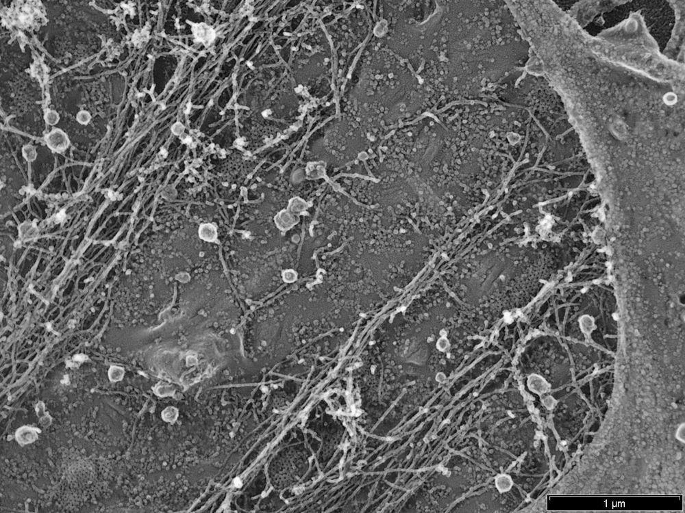
I am excited to share our first Pt replica EM images. It took us a little while, but now we have establish the unroofing, drying and Pt coating workflow 🎉 Great work by our postdoc Luis Wong Dilworth! The image below shows the cytosolic membrane leaflet of a fibroblast 👇
February 13, 2025 at 2:34 PM
I am excited to share our first Pt replica EM images. It took us a little while, but now we have establish the unroofing, drying and Pt coating workflow 🎉 Great work by our postdoc Luis Wong Dilworth! The image below shows the cytosolic membrane leaflet of a fibroblast 👇
Hello new followers 🖖 I like looking at the plasma membrane by high res microscopy. Here, you see a platinum replica EM of a fibroblast plasma membrane with clathrin dome (in the center) and caveolae (small invaginations on the side)
November 28, 2024 at 8:11 PM
Hello new followers 🖖 I like looking at the plasma membrane by high res microscopy. Here, you see a platinum replica EM of a fibroblast plasma membrane with clathrin dome (in the center) and caveolae (small invaginations on the side)
Lipid droplets for everyone! #fluorescencefriday

November 15, 2024 at 1:00 PM
Lipid droplets for everyone! #fluorescencefriday
Here are some more images of clathrin


October 7, 2023 at 4:23 PM
Here are some more images of clathrin
Caveolae‘s big brother : clathrin! Here we can see beautiful clathrin lattices deforming the plasma membrane lipids to a dome. Some of them will end up as spheres that can be internalized into the cell (this process is called endocytosis)

October 7, 2023 at 4:22 PM
Caveolae‘s big brother : clathrin! Here we can see beautiful clathrin lattices deforming the plasma membrane lipids to a dome. Some of them will end up as spheres that can be internalized into the cell (this process is called endocytosis)
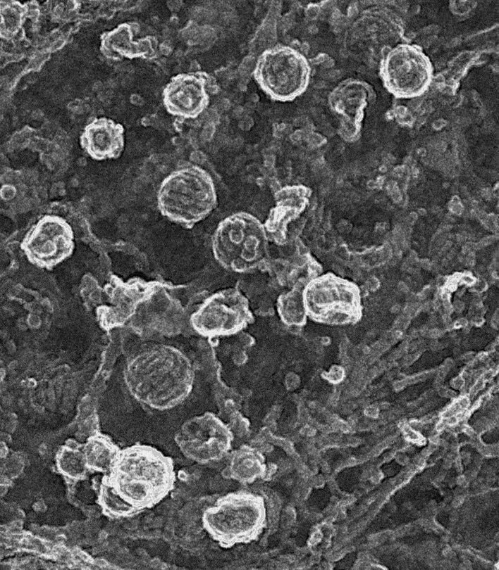
Caveolae are small plasma membrane invaginations (50-100 nm) found in many cell types. Here you can see the typical shape and coat structure of caveolae - in particular the cavin protein stripe coat is visible. (We are looking from the inside of a cell to the plasma membrane)
October 6, 2023 at 8:27 AM
Caveolae are small plasma membrane invaginations (50-100 nm) found in many cell types. Here you can see the typical shape and coat structure of caveolae - in particular the cavin protein stripe coat is visible. (We are looking from the inside of a cell to the plasma membrane)
Hello Blue sky, I like caveolae and lipid droplets

October 5, 2023 at 12:39 PM
Hello Blue sky, I like caveolae and lipid droplets