Development
@dev-journal.bsky.social
6K followers 580 following 490 posts
Development is a leading research journal in the field of developmental biology, covering stem cells, regeneration, evo-devo, epigenetics, morphogenesis and more. @biologists.bsky.social
Posts Media Videos Starter Packs
Pinned
dev-journal.bsky.social
Issue 18 is complete!

On the cover: Clones on Drosophila retina with activated Mbt kinase (pink), expanding Sdk protein (yellow) from tri- to bi-cellular adheren junctions & distorting the ommatidial lattice (cyan).

See Research Article by Gandhi et al.

journals.biologists.com/dev/article/...
Reposted by Development
biologists.bsky.social
Our first extraordinary biologist featured this week is Katherine Brown, @biologists.bsky.social Publishing Director and former @dev-journal.bsky.social Executive Editor, who has been closely involved with @prelights.bsky.social since its inception. #100biologists

@katherine-brown.bsky.social
The Company of Biologists 100 logo to the left and QR code to the right.

Portrait of Katherine Brown to the left, text to the right 100 extraordinary biologists 

Katherine Brown is the Company’s Publishing Director, having previously served as Development’s Executive Editor. There, she co-initiated the ‘From Stem Cells to Human Development’ meeting series, helping to launch the Company’s Journal Meetings. Katherine has also been closely involved with preLights since its inception.
dev-journal.bsky.social
To find out more about this work and the people behind it, we spoke to first author Isidora Rovic and corresponding author Andrea Juriscova, Associate Professor at the Lunenfeld-Tanenbaum Research Institute, Toronto, Canada.

journals.biologists.com/dev/article/...
First author Isidora Rovic (left) and corresponding author Andrea Jurisicova (right)
dev-journal.bsky.social
Read the #OA article “Acid sphingomyelinase is a gatekeeper of placental labyrinthine architecture and function” here:

journals.biologists.com/dev/article/...
dev-journal.bsky.social
Sphingolipid metabolism impacts placental processes

This Research Highlight showcases the work from Isidora Rovic, Andrea Jurisicova and colleagues:

journals.biologists.com/dev/article/...
Figure 4 - Placental defects in Smpd1−/−mice. (A) Morphology of gestational day (d) 17.5 wild-type (left) and Smpd1−/− placenta (right) at 2× magnification. (B) Total placental surface area was not significantly different between wild type and Smpd1−/−; however, the proportion of the junctional layer was increased and the proportion of labyrinth (Lab) was decreased in Smpd1−/− placentas (P<0.05; two-way ANOVA and Bonferroni post-hoc test). (C) Transmission electron micrographs of d17.5 wild-type and Smpd1−/− placenta interhemal membrane. (D) Maternal membrane space measurements are decreased in Smpd1−/− d15.5 and d17.5 placentas from six randomized rectangular sections (unpaired t-test). (E) Pseudo-colored transmission electron micrographs of d17.5 wild-type (top) and KO (bottom) interhemal membranes. MBS, maternal blood space; FC, fetal capillary; green pseudo-color indicates syncytiotrophoblast layer I (MBS facing); red pseudo-color indicates syncytiotrophoblast layer II (FC facing). (F) Interhemal thickness was significantly increased in KO labyrinth (unpaired t-test). (G) Expression of Mct1 (ST-I, green) and Mct4 (ST-II, red) immunofluorescence confocal microscopy in d17.5 wild-type and Smpd1−/− placentas. Mutant placentas exhibit focally elevated MCT1, as well as less defined and disorganized MCT4 staining. Nuclei are counterstained with DAPI (blue). (H) mRNA levels of Syna were significantly decreased in Smpd1−/− placentas (unpaired t-test). Individual data points represent individual placentas, obtained from at least two different dams. Letters indicate statistical significance (P<0.05). All data are mean±s.e.m. For details of the statistical test(s) used, see the Materials and Methods. Scale bars: 5 mm in A; 5 μm in C; 1 μm in E; 7 μm in G.
dev-journal.bsky.social
Pathway to Independence: a forecast for the future of developmental biology

In this Perspective, each of our 2025 Pathway to Independence Fellows discuss future outlooks for #DevBio and how their work will contribute to this exciting new era.

journals.biologists.com/dev/article/...
Picture of the title of the article and author list (2025 PI fellows) Chee Kiang Ewe, Max S. Farnworth, Anzy Miller, Joaquı́n Navajas Acedo, Marlies E. Oomen, Giulia Paci, Sonya A. Widen and Toshimichi Yamada
dev-journal.bsky.social
Also in Issue 18:

▪️4 Research highlights on cellular junctions, dendrite branching, teeth formation and gastruloid patterning
▪️3 People behind the paper interviews
▪️Transitions in development interview
▪️2 Perspectives
▪️Hypothesis on plant circadian genes

journals.biologists.com/dev/issue/15...
dev-journal.bsky.social
Issue 18 is complete!

On the cover: Clones on Drosophila retina with activated Mbt kinase (pink), expanding Sdk protein (yellow) from tri- to bi-cellular adheren junctions & distorting the ommatidial lattice (cyan).

See Research Article by Gandhi et al.

journals.biologists.com/dev/article/...
dev-journal.bsky.social
In preprints: gastruloids get (a)head

Wilhelmus A. Brouwer @wesselbrouwer.bsky.social and
Luca Braccioli @bracciolilab.bsky.social discuss the #preprint by Balaskas et al. "An advanced head-to-tail mouse embryo model with hypoxia-mediated neural patterning”

journals.biologists.com/dev/article/...
In Preprints: A new preprint curation initiative from Development
dev-journal.bsky.social
In preprints: gastruloids get (a)head

@wesselbrouwer.bsky.social and @bracciolilab.bsky.social discuss the #preprint by Balaskas et al. "An advanced head-to-tail mouse embryo model with hypoxia-mediated neural patterning”

journals.biologists.com/dev/article/...
dev-journal.bsky.social
To learn more about this story and the people behind it, we spoke to co-first authors @judithpineau.bsky.social and @jeromewongng.bsky.social, and corresponding author @thomasgregor.bsky.social, Head of Structure at @pasteur.fr & Professor at @princeton.edu

journals.biologists.com/dev/article/...
From left to right: co-first authors Judith Pineau and Jerome Wong-Ng, and corresponding author Thomas Gregor
dev-journal.bsky.social
Read the #OA article “Fine-tuning mechanical constraints reveals uncoupled patterning and gene expression programs in murine gastruloids” here:

journals.biologists.com/dev/article/...
dev-journal.bsky.social
Modifying mechanical constraint guides gastruloid patterning

This Research Highlight showcases the work from Judith Pineau @judithpineau.bsky.social, Jerome Wong-Ng @jeromewongng.bsky.social, Thomas Gregor @thomasgregor.bsky.social and colleagues:

journals.biologists.com/dev/article/...
Figure 2 - Gene expression patterning and transcriptional profiles in hydrogel-grown gastruloids. (A) Representative immunofluorescence images of gastruloids at 120 h. Gastruloids were grown in standard culture conditions (Ctrl, no hydrogel) or embedded in hydrogels with increasing concentrations (0.7 mM, 0.8 mM and 1.0 mM) at 96 h post-seeding. Nuclei (blue), BRA (purple) and SOX2 (yellow) are labeled. (B,C) Normalized expression profiles of SOX2 (B) and BRA (C) along the AP axis, demonstrating consistent expression patterns across conditions. (D) Principal component analysis of bulk RNA-seq experiments (three replicates), revealing clear separation between Ctrl and noCHI (red), a negative control for gastruloid formation, while hydrogel-embedded conditions cluster near Ctrl. (E) Number of significantly differentially expressed (DE) genes compared to Ctrl, showing substantial transcriptional changes in noCHI and minimal changes in hydrogel-embedded conditions (0.7 mM, 0.8 mM and 1.0 mM). Shown here are representative data and analysis for one experimental replicate. See Fig. S3 for two additional replicates.
dev-journal.bsky.social
Circadian clock genes perform dual roles as systemic oscillators and context-dependent regulators

A Hypothesis by Nozomu Takahashi and Motomu Endo that clock genes perform roles in both local regulation and systemic co-ordination during plant development.

journals.biologists.com/dev/article/...
Figure 2 - A conceptual model of the plant circadian system as a modular, dual-function regulatory network. This model builds upon the classical view of a unified, systemic oscillator (e.g. Somers, 1999; McClung, 2000) and its subsequent refinement into a complex regulatory network (e.g. Mas, 2005; Harmer, 2009; Pruneda-Paz and Kay, 2010) by proposing a module centric framework that resolves the functional uncoupling of rhythmicity and development. The model posits two parallel output pathways originating from organ-specific oscillators. Environmental inputs (left) entrain these oscillators, located in the leaf, shoot apex and root. First, these local oscillators function as network timekeepers, each generating a core circadian rhythm (wavy lines). The dashed arrows represent the contribution of each local oscillator to the systemic clock (bottom). This collective contribution establishes the systemic clock as a cohesive, emergent property that synchronizes the entire plant. Second, and in parallel, this model posits that specific modules of clock genes within each oscillator also function as direct, context-dependent regulators. They generate distinct output signals, which are then transmitted (shown as colored solid arrows below the wavy lines) to control key developmental processes. This regulatory pathway acts in parallel with the core timekeeping role of the clock components. This dual-function perspective explains how the plant circadian system can simultaneously maintain robust, whole-plant rhythmicity while executing highly specific, localized developmental programs.
dev-journal.bsky.social
Speaking of which, remember to save the date and register your interest for our 2026 Journal Meeting #HumanDev26 on 7th-9th September, co-organised with @wellcometrust.bsky.social-funded consortium Human Developmental Biology Initiative (HDBI)

www.biologists.com/meetings/dev...
Meeting Title - Human Development: Stem Cells, Models, Embryos. 
Date – 7th to 9th September 2026. 
Location – University of Warwick, UK. 
Organised by Development and the Human Developmental Biology Initiative (HDBI). 
Social media hashtag - #HumanDev26
dev-journal.bsky.social
From stem cells to human development: supporting a growing field

This Perspective from @katherine-brown.bsky.social reflects on the history of our journal meetings and their impact on both the developmental biology community and @biologists.bsky.social

journals.biologists.com/dev/article/...
‘From Stem Cells to Human Development’ over the years. (A,B) Official conference photos from the 2014 (A) and 2024 (B) meetings, both held at Wotton House in Surrey, UK. (C) Word cloud summarising feedback from meeting attendees at the 2014 event asked to describe their experience in four words. The largest and most immediately visible are: 'friendly atmosphere', 'interactive', 'inspirational', 'interesting', 'innovative', 'stimulating'. (D) Aggregate responses to questions from each post-event survey asking attendees to rate the meeting on a scale of 1-10 (orange bars) and whether they would recommend one of our events to a colleague. The recommendation score shows the percentage that responded ‘yes’ (green diamonds).
dev-journal.bsky.social
Also in Issue 17:

▪️4 Research highlights on cell fate bifurcation, axis determination, thermal stress and somitogenesis
▪️4 People behind the paper interviews
▪️Transitions in development interview
▪️2 Perspectives
▪️Review on human primordial germ cells

journals.biologists.com/dev/issue/15...
dev-journal.bsky.social
Our next #DevPres webinar focusses on development across scales with talks from Osvaldo Contreras @osvaldoics.bsky.social & Yinan Wan, chaired by Alex Eve @amjeve.bsky.social

📅 15 October, 09:00 BST (UTC+1)

Register here: us02web.zoom.us/webinar/regi...
Poster of Development presents... webinar on Wednesday 15 October at 09:00 BST (UTC+1). Chair: Alex Eve. Details of 2 speakers and talks: Osvaldo Contreras from the Victor Chang Cardiac Research Institute, talk titled 'OpenEMMU: A low-cost EdU multiplexing methodology for studying DNA replication and cell cycle dynamics’; then Yinan Wan from Biozentrum University of Basel, talk titled ‘Whole-embryo spatial transcriptomics: from fate to form’.
dev-journal.bsky.social
Issue 17 is complete!

On the cover: Image of three Hydra specimens. A wild-type Hydra (centre) and two transformants showing switch of foot (pink) to tentacle (yellow) (right) and tentacles to feet (left).

See Research Article by Ferenc et al.

journals.biologists.com/dev/article/...
dev-journal.bsky.social
To learn more about this work and the people behind it, we spoke to first author Hannah Brown and corresponding author Sean Ryder @seanryderphd.bsky.social, Professor and Vice Chair for Outreach, Biochemistry and Molecular Biotechnology @umasschan.bsky.social

journals.biologists.com/dev/article/...
Sean Ryder and research group, including first author Hannah Brown. Picture shows seven people with their hands up and/or doing jazz hands in a hallway.
dev-journal.bsky.social
Read the article "The mex-3 3′ untranslated region is essential for reproduction during temperature stress" here:

journals.biologists.com/dev/article/...
dev-journal.bsky.social
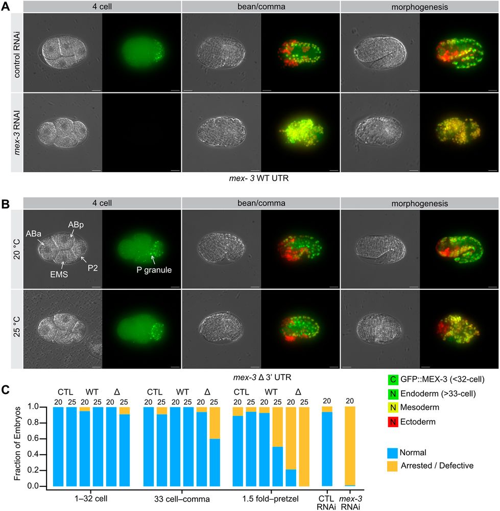
MEX-3’s untranslated region helps worms beat the heat

This Research Highlight showcases the work from Sean Ryder (@seanryderphd.bsky.social) and colleagues:

journals.biologists.com/dev/article/...
Figure 5: Germ layer patterning defects in the mex-3 Δ3′UTR mutant. (A) mex-3 RNAi results in dead embryos with a large expansion of muscle lineage cells (mesoderm, yellow). Control animals develop normally, with all three germ layers present and appropriately patterned. Cytoplasmic (C) green represents GFP::MEX-3 protein, nuclear (N) green indicates endoderm, nuclear (N) yellow indicates mesoderm and nuclear (N) red indicates ectoderm. (B) At elevated temperature, the mex-3 Δ3′UTR mutants form all three germ layers, but patterning appears defective with a moderate expansion of muscle lineage cells (yellow). (C) Fraction of embryos that appear normal versus abnormal binned as in Fig. 4. The genotype and the temperature of growth are shown. CTL is a control for the germ layer background (strain OD1854). All sample sizes and statistical test outcomes are listed in Table S3. Scale bars: 10 μm.
dev-journal.bsky.social
Transitions in development – an interview with Mijo Šimunović
We caught up with Mijo Šimunović from @columbiauniversity.bsky.social to learn more about Mijo's transition to becoming a group leader and Mijo's insights on the growing need for cross-disciplinary research: doi.org/10.1242/dev....
Portrait of Mijo Šimunović
dev-journal.bsky.social
To learn more about this work and the people behind it, we spoke to first author Zoe Chen, and the corresponding author, Tyler Square, Assistant Professor at the University of Florida, USA: doi.org/10.1242/dev....
Tyler Square (left) and Zoe Chen (right)
dev-journal.bsky.social
Wrong place, right time: Eda drives ectopic teeth formation in fish
This Research Highlight showcases the work from Tyler Square and colleagues: journals.biologists.com/dev/article/...
Eda overexpression causes precocious tooth differentiation in sticklebacks. (A,B) at 9 dpf, Eda OE fish (n=9) demonstrated precocious oral tooth germs relative to control fish (n=10). Images show eGFP from the dlx2b tooth reporter and mCherry from the OE transgene. A’ and B’ show the fish indicated in A and B in greater detail. Black arrowheads indicate pioneer tooth germs. (C) at 12 dpf in WT, pairs of germs are observed on the premaxilla and dentary (black arrowheads), similar to the condition seen in the 9 dpf Eda OE fish, suggesting these represent accelerated endogenous teeth. Sale bars=100 μm.