Houart Lab
@houartlab.bsky.social
280 followers
240 following
20 posts
Our lab studies fundamental mechanisms in early neurodevelopment and neurodegenerative diseases | Centre for Developmental Neurobiology @ King's College London | Satellite lab @ The Francis Crick Institute
Posts
Media
Videos
Starter Packs
Reposted by Houart Lab
Houart Lab
@houartlab.bsky.social
· Jul 18
Reposted by Houart Lab
Reposted by Houart Lab

Reposted by Houart Lab


Reposted by Houart Lab
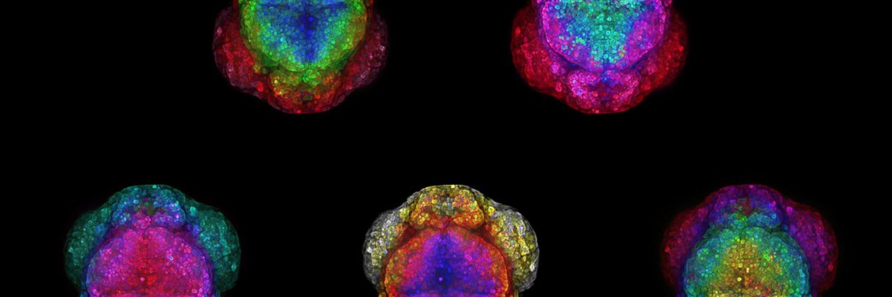
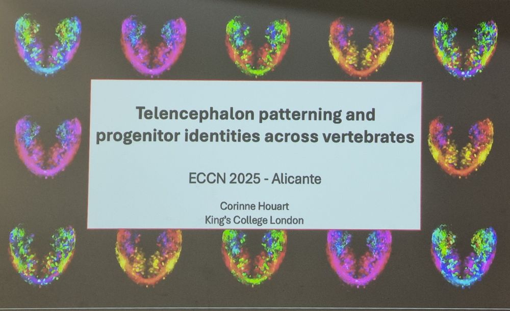
Houart Lab
@houartlab.bsky.social
· Jun 3
Reposted by Houart Lab
Reposted by Houart Lab


Reposted by Houart Lab
Reposted by Houart Lab
Reposted by Houart Lab
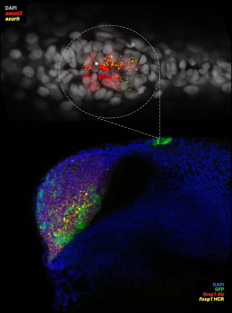
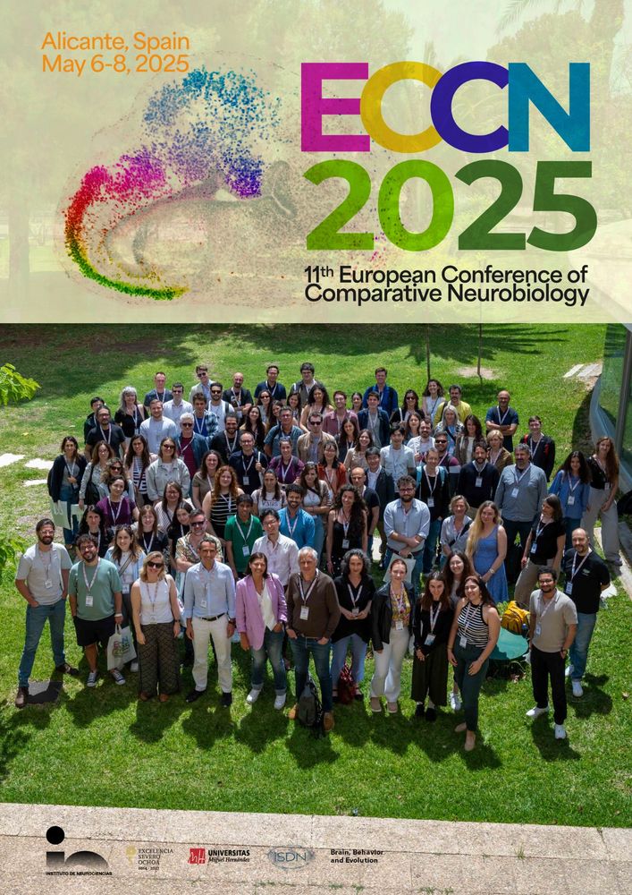
Houart Lab
@houartlab.bsky.social
· May 6