ISPH 2025
@isph2025.bsky.social
36 followers 20 following 60 posts
7th International Symposium on Palaeohistology (ISPH), Brisbane, QLD, Australia - June 2025 http://www.isph2025.weebly.com
Posts Media Videos Starter Packs
Pinned
isph2025.bsky.social
#isph2025 group photo!
Reposted by ISPH 2025
gricabiopastudc.bsky.social
Our #BIOpast team was there! Huge thanks to the organizers for such an amazing experience 🥳 #PaleoGalicia #Paleomeeting
isph2025.bsky.social
#isph2025 group photo!
isph2025.bsky.social
#isph2025 group photo!
Reposted by ISPH 2025
draliceclement.bsky.social
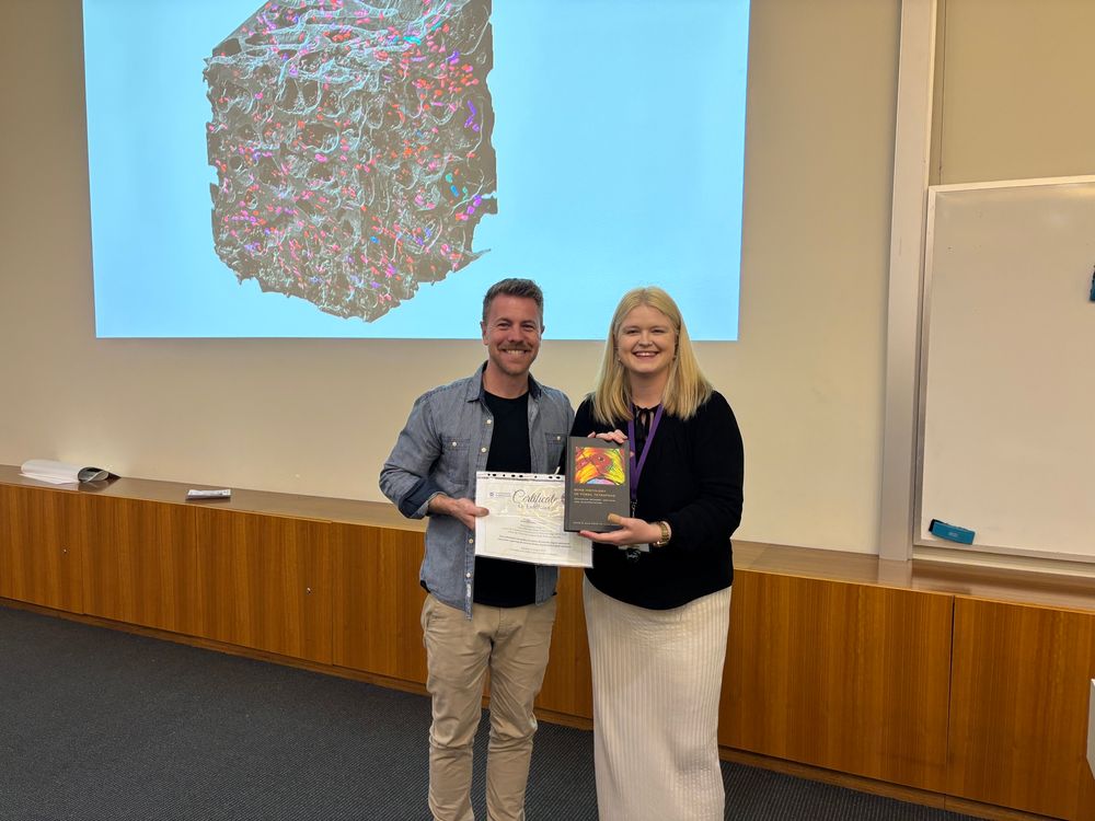
Congratulations to M. Ramon Fritzen, PhD candidate at Flinders University, for winning 2nd place in the @isph2025.bsky.social image competition using a @ansto.bsky.social MCT scan of (Gogo) fossil lungfish bone!
isph2025.bsky.social
Marcos Ramon Fritzen won joint 2nd place of our palaeohistology image competition with his image titled "A Deep Time Genomic Window: 3D Visualization of Fossil Lungfish Osteocyte Lacunae" #isph2025
Reposted by ISPH 2025
palaeo-prof.bsky.social
@isph2025.bsky.social Thanks to @justyna Miszkiewicz and team for a fabulous ISPH meeting! @paleohistology
Reposted by ISPH 2025
boneremodelling.bsky.social
Congratulations to our postdoc @bonehistopath.bsky.social for winning 2nd place of the #isph2025 image competition!
isph2025.bsky.social
@bonehistopath.bsky.social won joint 2nd place of our palaeohistology image competition with her image titled "Desert Landscape" #isph2025
isph2025.bsky.social
Winners of our palaeohistology image competition #isph2025

1st place: Professor Tim Bromage
Joint 2nd: @bonehistopath.bsky.social & Marcos Ramon Fritzen
isph2025.bsky.social
It marks a methodological shift from traditional descriptive histology to a quantitative and predictive approach that connects fossil microstructure with the genomic history of vertebrates.
isph2025.bsky.social
As part of a growing digital dataset, this reconstruction contributes to identifying evolutionary trends in lungfishes through time.
isph2025.bsky.social
Rather than focusing solely on bone morphology, it enables the quantification of cellular features as big data, allowing statistical analysis and observation of trends in volume and shape variation.
isph2025.bsky.social
This work highlights the integration of high-resolution imaging, artificial intelligence segmentation, and large-scale data generation in palaeontology.
isph2025.bsky.social
Applying this principle to fossil taxa offers new insight into genome evolution in early sarcopterygians, linking histological structures to deep-time genomic patterns.
isph2025.bsky.social
Using a deep learning-based segmentation, individual osteocyte lacunae were digitally isolated from fossilized bone in unprecedented detail. Osteocyte lacuna volume is a recognized proxy for genome size in living vertebrates.
isph2025.bsky.social
This image presents the first 3D visualization of osteocyte lacunae in a 382 million-year-old lungfish from Australia, Chirodipterus australis, scanned at ANSTO with a voxel size of 0.71 microns.
isph2025.bsky.social
Marcos Ramon Fritzen won joint 2nd place of our palaeohistology image competition with his image titled "A Deep Time Genomic Window: 3D Visualization of Fossil Lungfish Osteocyte Lacunae" #isph2025
isph2025.bsky.social
This image captures a fleeting moment in that ongoing war--a testament to both the resilience and vulnerability of the human body.
isph2025.bsky.social
Under the lens of confocal laser scanning microscopy, this microscopic battlefield is revealed in striking detail: a dry, hostile landscape where infection and healing vie for control. Will the bone recover, or will the pathogens prevail?
isph2025.bsky.social
Pits, hollows, mountains, and cliffs--not features of an ancient desert, but signs of the body’s struggle against disease. Infected and inflamed, bone becomes a dynamic terrain, reshaped by periosteal growth as the body fights to repair itself.
isph2025.bsky.social
@bonehistopath.bsky.social won joint 2nd place of our palaeohistology image competition with her image titled "Desert Landscape" #isph2025
isph2025.bsky.social
Preferred orientations inform on the nature of forces encountered in habitual use of Lucy’s femur in life. A montage of the entire cross section acquired this way revealed a pattern of preferred orientations that are consistent with the loading history of a bipedal human. Field width 600 μm.
isph2025.bsky.social
In this real color image, green represents collagen oriented perpendicular to the image, and white represents collagen transversely oriented in Haversian systems.
isph2025.bsky.social
This novel illuminating method generates autofluorescence of the bone matrix, and provides image contrast regarding degrees of birefringent collagen fiber orientations.
isph2025.bsky.social
After three years of innovation and engineering, I went to Addis Ababa, and with permission from the Ethiopia National Museum, a fracture across the mid-shaft femur was imaged using circularly polarized ultraviolet light.
isph2025.bsky.social
To achieve the task of researching Lucy’s skeletal microanatomy in the face of these constraints, it was necessary to build a portable confocal scanning optical microscope and perform optical thin section paleohistology.
isph2025.bsky.social
There exists a class of objects that are rigorously defended from thin section paleohistology and that cannot travel to an imaging workstation for study. One such object is the famous “Lucy”, Australopithecus afarensis, discovered from fossil bearing deposits at Hadar, Ethiopia, 3.0 m.y.
isph2025.bsky.social
We are pleased to announce the winner of our palaeohistology image competition is Professor Tim Bromage with his image titled "Lucy Fiber Orientation" #isph2025