Journal of Anatomy
@journalofanatomy.bsky.social
460 followers
240 following
51 posts
Official journal of the Anatomical Society (@anat_soc). We improve understanding of anatomy through analysis of structure, function, development and evolution: https://onlinelibrary.wiley.com/journal/14697580
Posts
Media
Videos
Starter Packs


Robyn Grant
@robyngrant.bsky.social
· Jun 11
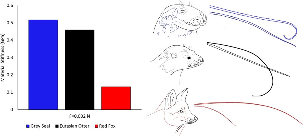
Determining modulus of elasticity using finite element analysis and non‐destructive testing: Are aquatic animal whiskers stiffer?
The values of the modulus of elasticity (E) for each species whiskers were obtained when the FE model was aligned to the maximum displacement values of the experiment. Grey seal E values were larger ...
onlinelibrary.wiley.com