
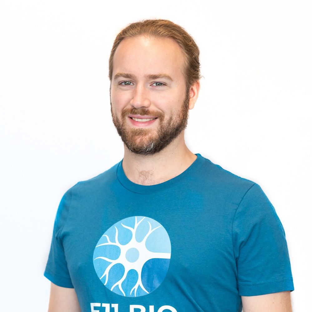
Josiah Passmore
@jpassmore.bsky.social
370 followers
550 following
25 posts
Making cells run in circles, and gels with squares in them.
Automated optogenetics, Smart microsopy, Expansion microscopy, GelMap.
Postdoc @ Utrecht University | visualise.bio.
Posts
Media
Videos
Starter Packs
Pinned

Reposted by Josiah Passmore
Reposted by Josiah Passmore

Reposted by Josiah Passmore
Reposted by Josiah Passmore

Reposted by Josiah Passmore
Reposted by Josiah Passmore
Reposted by Josiah Passmore
Malte Kuehl
@maltekuehl.com
· Jul 18
Reposted by Josiah Passmore
Reposted by Josiah Passmore

Reposted by Josiah Passmore
Reposted by Josiah Passmore


Reposted by Josiah Passmore
Reposted by Josiah Passmore
Reposted by Josiah Passmore
Reposted by Josiah Passmore
Reposted by Josiah Passmore


Reposted by Josiah Passmore
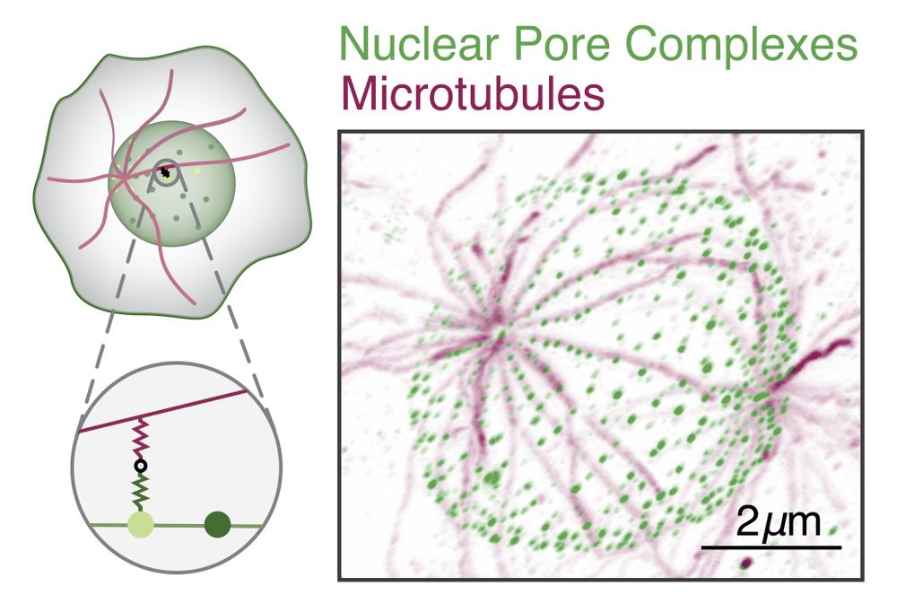
Malina Iwanski
@mkiwanski.bsky.social
· Feb 9

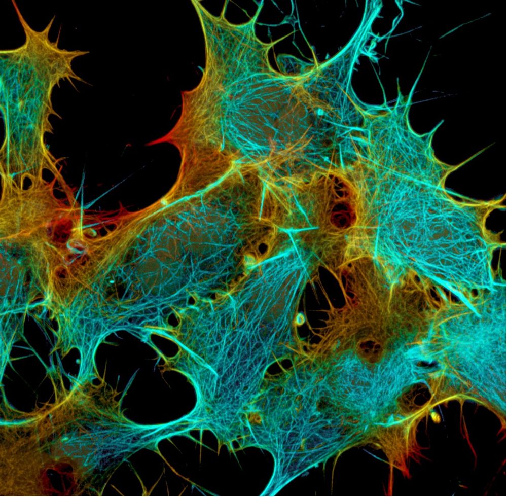
Polarity reversal of stable microtubules during neuronal development
Neurons critically depend on long-distance transport orchestrated by motor proteins walking over their highly asymmetric microtubule cytoskeleton. These microtubules are organized uniformly in axons w...
www.biorxiv.org

Reposted by Josiah Passmore
Reposted by Josiah Passmore