Jay R. Seltzer, MD
@jrseltzer.bsky.social
610 followers 110 following 120 posts
Medical Director Urine Microscopy Laboratory Missouri Baptist Medical Center St. Louis Missouri USA
Posts Media Videos Starter Packs
jrseltzer.bsky.social
Oval fat body and acanthocyte - brightfield with SM stain - in preserved urine sample from a case of GPA, stored at room temperature for 2.5 years! #UrineMicroscopy #UrinarySediment
jrseltzer.bsky.social
Calcium oxalate dihydrate crystals - brightfield and darkfield, unstained - from pt of @nephrothaniel.bsky.social - #UrineMicroscopy #UrinarySediment
jrseltzer.bsky.social
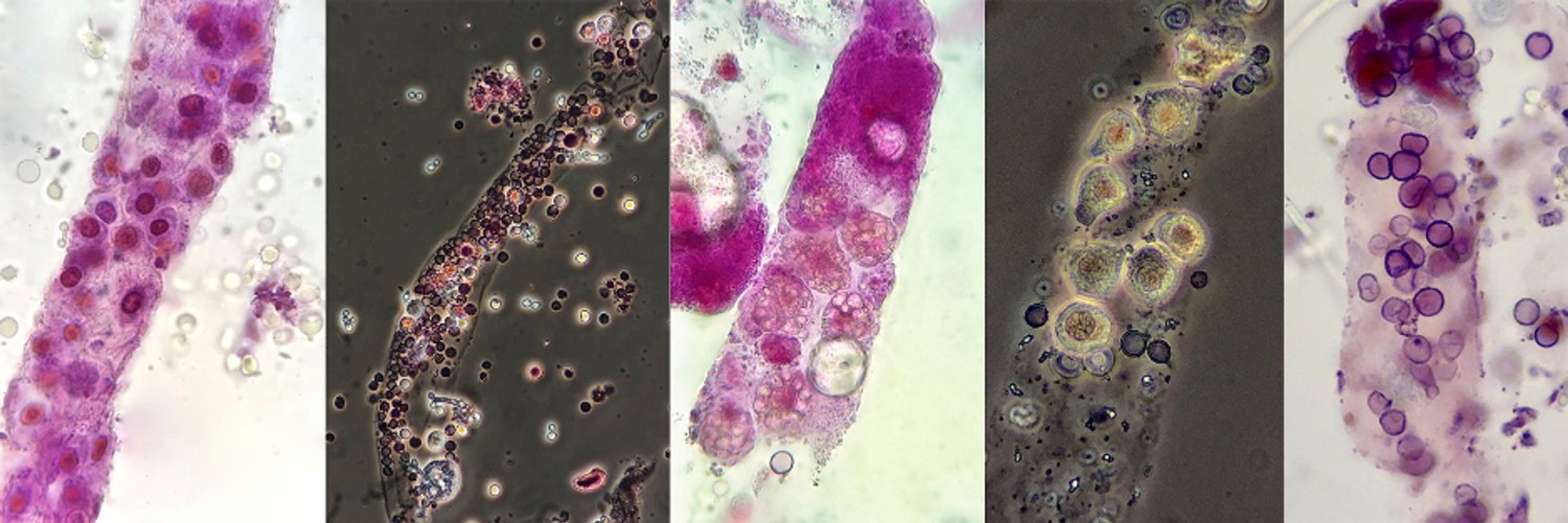
RBC cast - brightfield, unstained - from patient with hydralazine-induced vasculitis #UrineMicroscopy #UrinarySediment
jrseltzer.bsky.social
RBC's within waxy cast - brightfield and darkfield, unstained - from patient with hydralazine-induced vasculitis #UrineMicroscopy #UrinarySediment
jrseltzer.bsky.social
RBC cast - brightfield, with SM stain - from patient with hydralazine-induced vasculitis #UrineMicroscopy #UrinarySediment
jrseltzer.bsky.social
WBC cast - brightfield and darkfield, with SM stain - from patient with hydralazine-induced vasculitis #UrineMicroscopy #UrinarySediment
jrseltzer.bsky.social
Acanthocyte - phase contrast (original mag x400) - from patient with hydralazine-induced vasculitis #UrineMicroscopy #UrinarySediment
jrseltzer.bsky.social
RBC cast - brightfield with SM stain - from patient with MSSA bacteremia and osteomyelitis - #UrineMicroscopy #UrinarySediment
jrseltzer.bsky.social
RBC cast - brightfield with SM stain - from new case of MPA - #UrineMicroscopy #UrinarySediment
jrseltzer.bsky.social
Very long RBC cast - darkfield with SM stain - from new case of MPA - #UrineMicroscopy #UrinarySediment
jrseltzer.bsky.social
RBC casts - brightfield with SM stain - from new case of MPA - #UrineMicroscopy #UrinarySediment
jrseltzer.bsky.social
Acanthocyte - darkfield, unstained - cropped from original x1000 - from case of suspected hydralazine-induced vasculitis - #UrineMicroscopy #UrinarySediment
jrseltzer.bsky.social
RBC casts - brightfield with SM stain - from case of suspected hydralazine-induced vasculitis - #UrineMicroscopy #UrinarySediment
jrseltzer.bsky.social
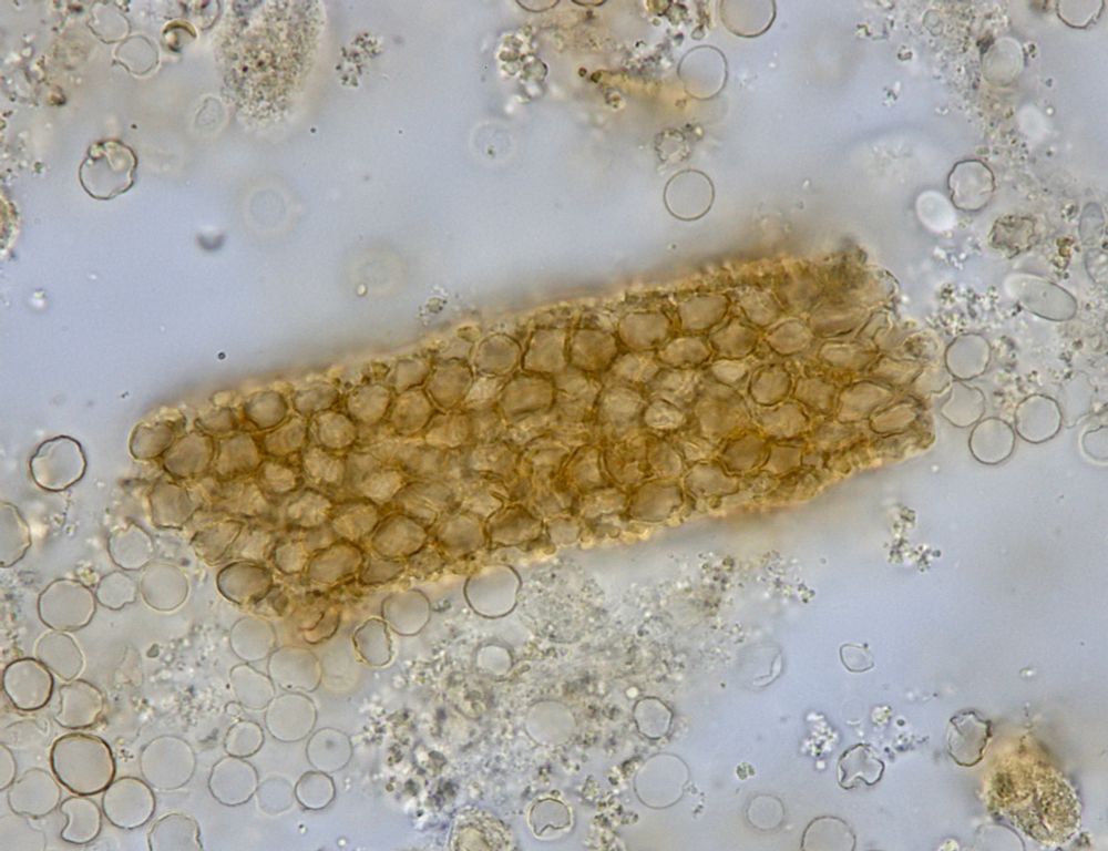
Vacuolar cast (which probably also contains some RBC's) - brightfield with SM stain - from patient with AKI and suspected hydralazine-induced vasculitis #UrineMicroscopy #UrinarySediment
Reposted by Jay R. Seltzer, MD
juancarlosqvelez.bsky.social
Interested in learning more about the art of microscopic examination of the urinary sediment and its clinical applications? @jrseltzer.bsky.social @swethakanduri.bsky.social and I will be running a Hands-On Workshop at KidneyCon this year. Come join us! @kidneycon.bsky.social May 1-3. Register ASAP
jrseltzer.bsky.social
New #UrinarySediment and #UrineMicroscopy case studies in @akdhjournal

Use the link below but be sure to view as a pdf - the online version is not formatted correctly

kwnsfk27.r.eu-west-1.awstrack.me/L0/https:%2F...
jrseltzer.bsky.social
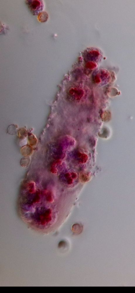
Young male presented with DVT and PE. Found to have UPC 8 gm/gm, serum albumin 2.2, and creatinine 2.3 - #UrineMicroscopy showed this mixed cellular cast containing RBC's, tubular epithelial cells, and WBC's - brightfield with SM stain - #UrinarySediment
jrseltzer.bsky.social
Vacation's over and I'm back in the lab....
Young male presented with DVT and PE. Found to have UPC 8 gm/gm and serum albumin 2.2 and creatinine 2.3 - #UrineMicroscopy showed RBC casts with fibrinous matrix as well as a few lipid casts - brightfield with SM stain - #UrinarySediment
jrseltzer.bsky.social
WBC cast - brightfield with SM stain - from patient with recurrent UTI's s/p abx rx over 3 weeks - #UrineMicroscopy #UrinarySediment
jrseltzer.bsky.social
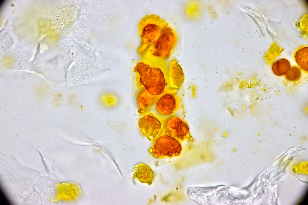
RBC cast - brightfield, unstained - from patient with IRGN - #UrineMicroscopy #UrinarySediment
jrseltzer.bsky.social
RBC's in a waxy cast - brightfield, unstained - from patient with IRGN - #UrineMicroscopy #UrinarySediment
jrseltzer.bsky.social
RBC cast with fibrinous matrix - brightfield with SM stain - from patient with AKI and chronic Staph. osteomyelitis - biopsy showed an IgA dominant IRGN pattern #UrinarySediment #UrineMicroscopy