Silas Busch
@neurosilas.bsky.social
170 followers
160 following
12 posts
Postdoc | Rockefeller University
Neurobiology PhD | UChicago
Works on: Dendrites, cerebellar circuits, comparative anatomy, fruit fly learning+memory
Dabbles in: Science art, philosophy, poetry, plants
Posts
Media
Videos
Starter Packs
Silas Busch
@neurosilas.bsky.social
· Apr 22

Reposted by Silas Busch
Blake Richards
@tyrellturing.bsky.social
· Apr 22

Cerebellum instructs plasticity in the mouse primary somatosensory cortex
Sensory experiences map onto distributed neural networks and may activate plasticity processes that in some brain areas are supervised by instructive signals. What remains unknown, however, is if such...
www.biorxiv.org
Silas Busch
@neurosilas.bsky.social
· Apr 18
Silas Busch
@neurosilas.bsky.social
· Apr 18
Silas Busch
@neurosilas.bsky.social
· Apr 18

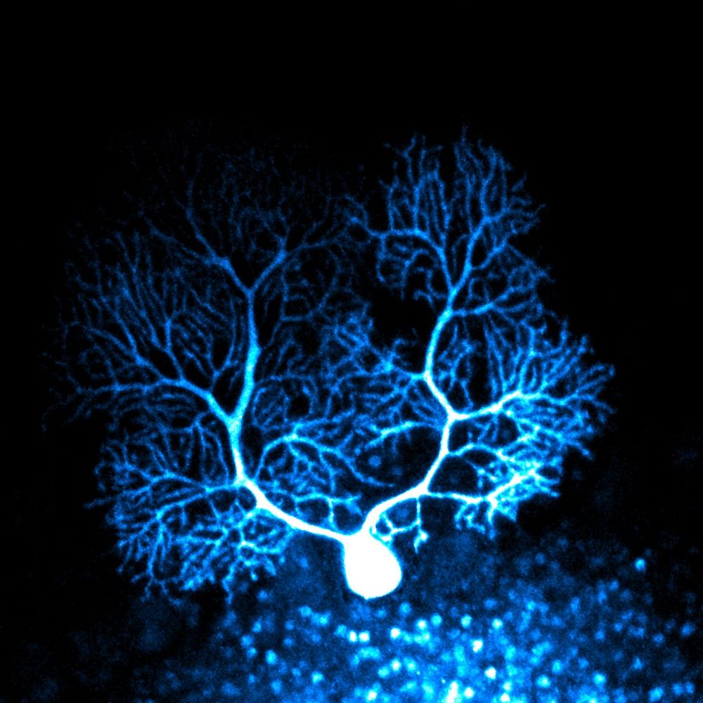
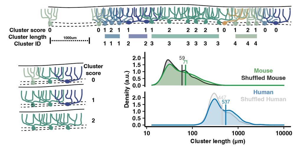
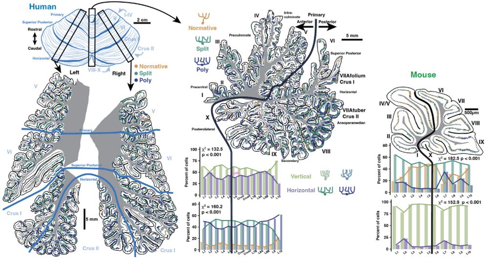
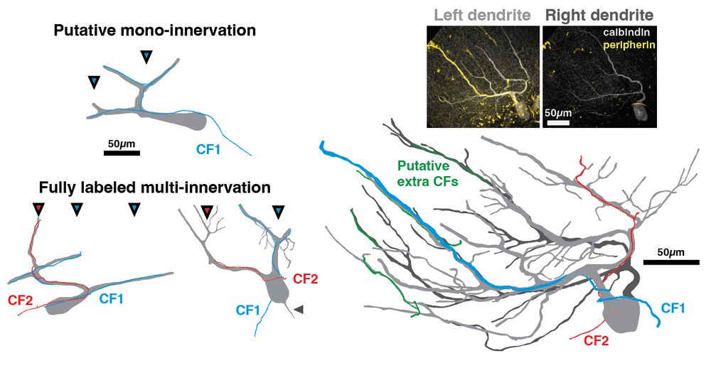
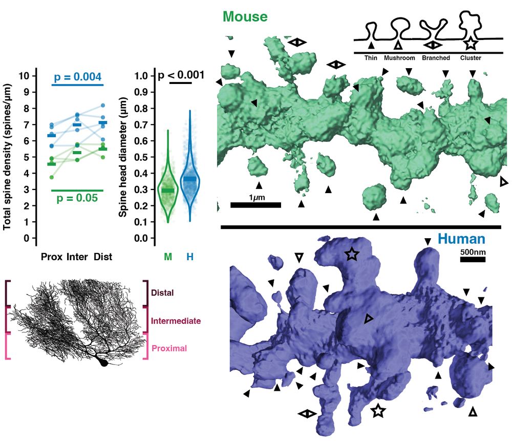
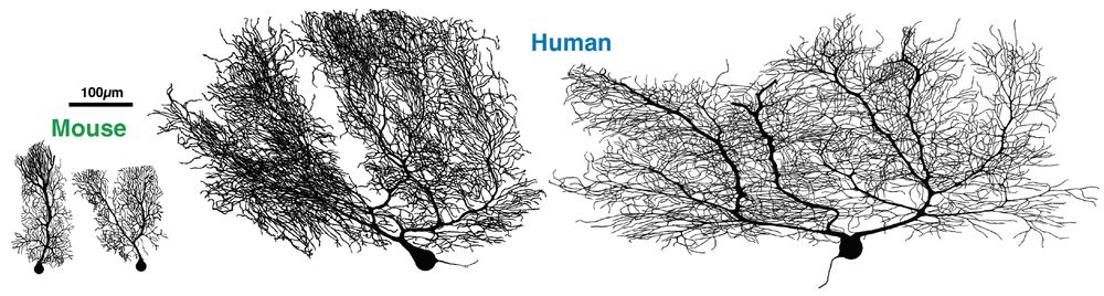
Non-allometric expansion and enhanced compartmentalization of Purkinje cell dendrites in the human cerebellum
A comparative study of Purkinje dendrite morphology, input arrangement, and regional subtype distribution shows human cells evade constraint by cortical thickness to be both quantitatively and qualita...
elifesciences.org
Silas Busch
@neurosilas.bsky.social
· Sep 11

Silas Busch
@neurosilas.bsky.social
· Oct 31