


Brian Kelch
@briankelch.bsky.social
My lab at UMass Chan Medical School studies virus assembly and DNA replication/repair using structural biology, biophysics, and biochemistry. Habitual Line-Stepper.
umassmed.edu/kelchlab
umassmed.edu/kelchlab
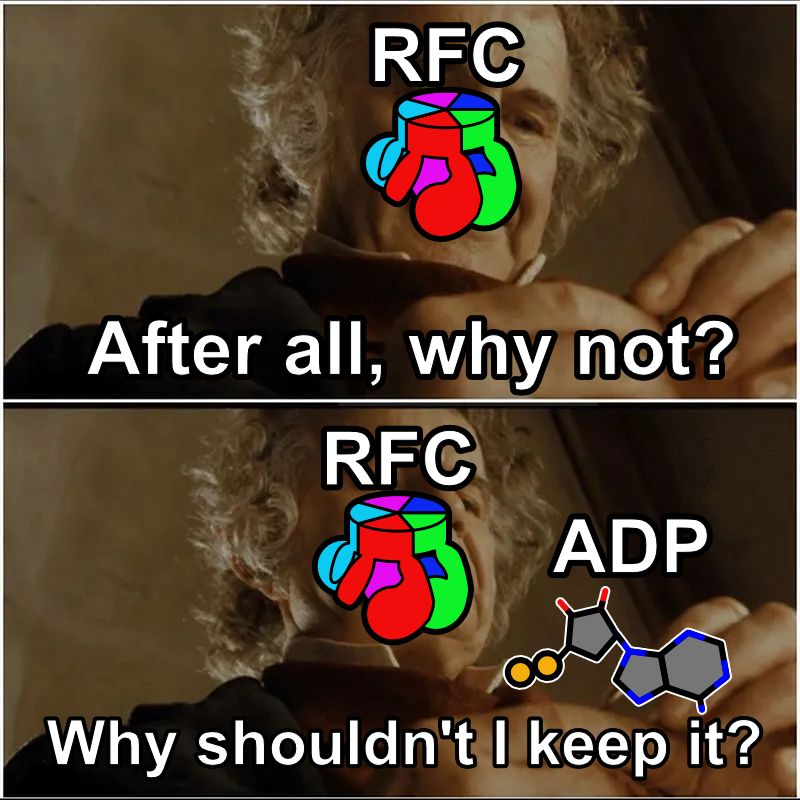
Every day we make enough DNA to go to the moon and back. So, all our DNA replication enzymes work fast, right? In our latest preprint, we show that a key enzyme, the clamp loader ATPase RFC, releases ADP much slower than it loads PCNA. What gives? (1/16)
doi.org/10.1101/2025...
doi.org/10.1101/2025...
July 7, 2025 at 7:06 PM
Every day we make enough DNA to go to the moon and back. So, all our DNA replication enzymes work fast, right? In our latest preprint, we show that a key enzyme, the clamp loader ATPase RFC, releases ADP much slower than it loads PCNA. What gives? (1/16)
doi.org/10.1101/2025...
doi.org/10.1101/2025...
Reposted by Brian Kelch
Hurray, it is finally out! Meet bacteriophage Bxb1 - the subject of my first full-phage cryo-EM study. My structures are beautifully complemented by the Park Lab’s cryo-ET analysis, shedding light on mycobacteriophage structural changes during infection.
www.sciencedirect.com/science/arti...
www.sciencedirect.com/science/arti...
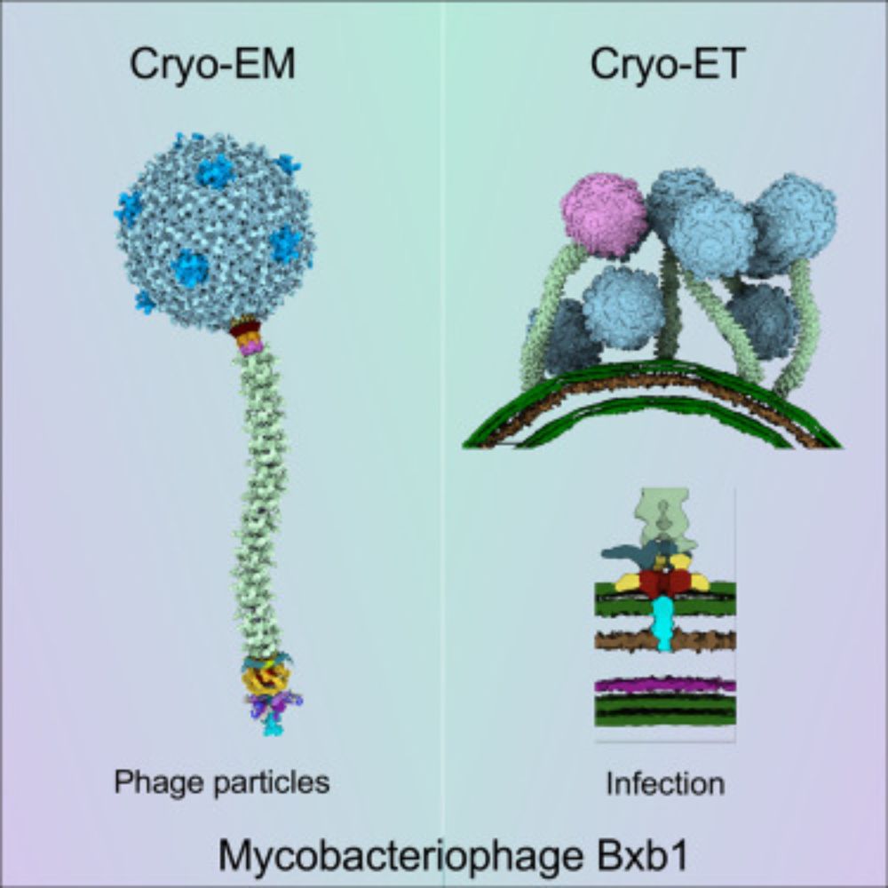
Structure and infection dynamics of mycobacteriophage Bxb1
Mycobacteriophage Bxb1 is a well-characterized virus of Mycobacterium smegmatis with double-stranded DNA and a long, flexible tail. Mycobacteriophages…
www.sciencedirect.com
April 16, 2025 at 12:50 PM
Hurray, it is finally out! Meet bacteriophage Bxb1 - the subject of my first full-phage cryo-EM study. My structures are beautifully complemented by the Park Lab’s cryo-ET analysis, shedding light on mycobacteriophage structural changes during infection.
www.sciencedirect.com/science/arti...
www.sciencedirect.com/science/arti...
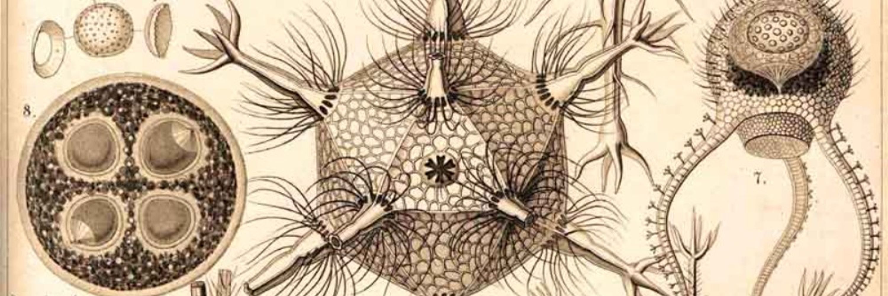
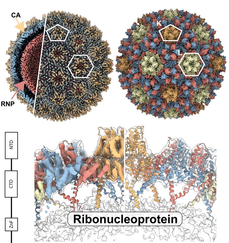
Did you know that there is so much DNA packed inside a phage capsid that the pressure is 10X higher than in a bottle of champagne? In our latest preprint, we wondered how that is contained in a phage that lives at extremely high temperatures. /1
www.biorxiv.org/content/10.1...
www.biorxiv.org/content/10.1...

April 18, 2025 at 2:09 PM
Did you know that there is so much DNA packed inside a phage capsid that the pressure is 10X higher than in a bottle of champagne? In our latest preprint, we wondered how that is contained in a phage that lives at extremely high temperatures. /1
www.biorxiv.org/content/10.1...
www.biorxiv.org/content/10.1...
I'm a little late to the game, but I'm happy to announce that our paper with my @umasschan.bsky.social colleagues in the Travis Thomson lab has now been published in @plosbiology.org! Thanks to all involved on this fun collaboration!

#Transposons are parasitic genetic elements that act as raw material for evolution of new functions. @briankelch.bsky.social &co show that the capsid of the #virus-derived retrotransposon Copia mediates synaptic plasticity at the #Drosophila neuromuscular junction 🧪 @plosbiology.org plos.io/3X71IDa
April 18, 2025 at 2:02 PM
I'm a little late to the game, but I'm happy to announce that our paper with my @umasschan.bsky.social colleagues in the Travis Thomson lab has now been published in @plosbiology.org! Thanks to all involved on this fun collaboration!
Reposted by Brian Kelch
Fun primer with @ForeverYHChang highlighting a lovely Travis Thomson lab paper!
Did animals co-opt viral gag for intercellular (trans synaptic) communication? Or did viruses co-opt retrotransposon gags that animals had previously co-opted for intercellular use?
Did animals co-opt viral gag for intercellular (trans synaptic) communication? Or did viruses co-opt retrotransposon gags that animals had previously co-opted for intercellular use?
Yung-Heng Chang & @joshdubnau.bsky.social explore a @plosbiology.org study revealing that the #retrotransposon gag protein of Copia forms virus-like capsids that transfer its own RNA across the fly neuromuscular junction to regulate #synapse formation 🧪 Paper: plos.io/3X71IDa Primer: plos.io/4i6rzmC

February 20, 2025 at 12:49 PM
Fun primer with @ForeverYHChang highlighting a lovely Travis Thomson lab paper!
Did animals co-opt viral gag for intercellular (trans synaptic) communication? Or did viruses co-opt retrotransposon gags that animals had previously co-opted for intercellular use?
Did animals co-opt viral gag for intercellular (trans synaptic) communication? Or did viruses co-opt retrotransposon gags that animals had previously co-opted for intercellular use?
Reposted by Brian Kelch
Congratulations to Jenna Whalen on her Nature Cancer paper out today-a distinct take on nicks and their toxicity! rdcu.be/d60Td
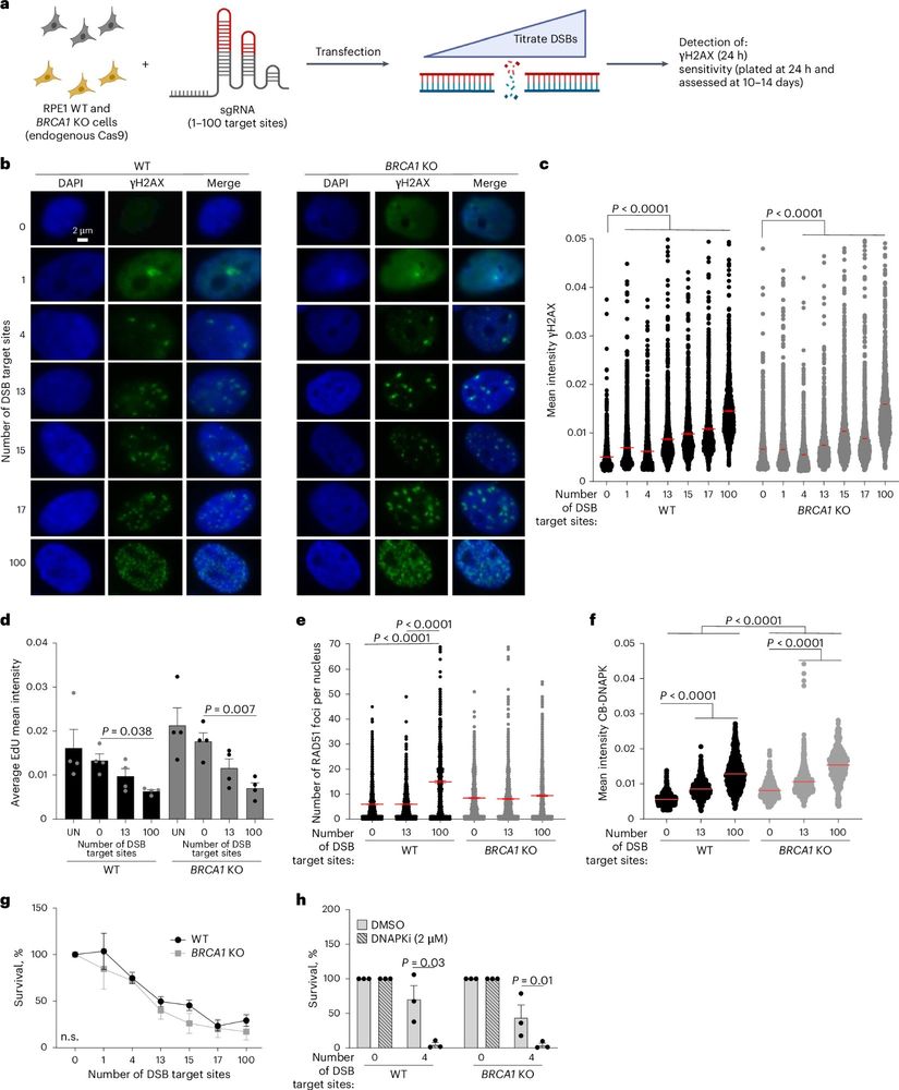
Targeting BRCA1-deficient PARP inhibitor-resistant cells with nickases reveals nick resection as a cancer vulnerability
Nature Cancer - Whalen et al. report that increased DNA end resection in BRCA-deficient, PARP inhibitor-resistant cancers leads to increased sensitivity to DNA nicks, limiting tumor formation in...
rdcu.be
January 21, 2025 at 5:26 PM
Congratulations to Jenna Whalen on her Nature Cancer paper out today-a distinct take on nicks and their toxicity! rdcu.be/d60Td
Reposted by Brian Kelch

Looking for TT faculty job? At #ASHG23? We’re recruiting! We = Genomics and Comp Bio dept at UMass Chan Medical School in Worcester, MA (not Boston but near Boston)
academicjobsonline.org/ajo/jobs/25641
pop genomics, imaging, stat. genetics, machine learning aka cool science w big data.
academicjobsonline.org/ajo/jobs/25641
pop genomics, imaging, stat. genetics, machine learning aka cool science w big data.
University of Massachusetts Chan Medical School, Department of Genomics and Computational Biology
Full service online faculty recruitment and application management system for academic institutions worldwide. We offer unique solutions tailored for academic communities.
academicjobsonline.org
November 2, 2023 at 5:06 PM
Looking for TT faculty job? At #ASHG23? We’re recruiting! We = Genomics and Comp Bio dept at UMass Chan Medical School in Worcester, MA (not Boston but near Boston)
academicjobsonline.org/ajo/jobs/25641
pop genomics, imaging, stat. genetics, machine learning aka cool science w big data.
academicjobsonline.org/ajo/jobs/25641
pop genomics, imaging, stat. genetics, machine learning aka cool science w big data.
Reposted by Brian Kelch
Hey, senior PhD students working on RNA and moving toward leading independent labs: Apply for the Maraganore Early Independence Fellow program at the UMass Chan RNA Therapeutics Institute and come work with us! www.linkedin.com/posts/angela...
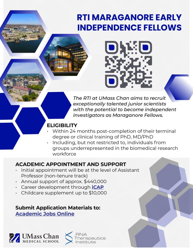
Angela Messmer-Blust on LinkedIn: #diversityinstem #underrepresentedinscience #rnabiology…
I am unbelievably excited and proud to launch the RNA Therapeutic Institute's new Maraganore Early Independence Fellows Program at UMass Chan Medical School…
www.linkedin.com
September 19, 2024 at 7:38 PM
Hey, senior PhD students working on RNA and moving toward leading independent labs: Apply for the Maraganore Early Independence Fellow program at the UMass Chan RNA Therapeutics Institute and come work with us! www.linkedin.com/posts/angela...
Reposted by Brian Kelch
Systems and synthetic biologists, we are recruiting at the department of Systems Biology at the @UMassChan. Position is open to faculty at all levels.
http://academicjobsonline.org/ajo/jobs/25423
http://academicjobsonline.org/ajo/jobs/25423
University of Massachusetts Chan Medical School, Departme...
Full service online faculty recruitment and application m...
academicjobsonline.org
November 23, 2024 at 7:28 AM
Systems and synthetic biologists, we are recruiting at the department of Systems Biology at the @UMassChan. Position is open to faculty at all levels.
http://academicjobsonline.org/ajo/jobs/25423
http://academicjobsonline.org/ajo/jobs/25423
Reposted by Brian Kelch
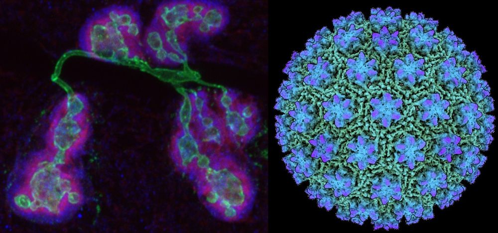
Over 4 years in the making, I am extremely happy to share our pre-print on the “In-cell structure and snapshots of copia retrotransposons in intact tissue”. We used cryo-ET to resolve the copia capsid structure to subnanometer resolution inside cells: www.biorxiv.org/content/10.1...
February 24, 2024 at 9:37 AM
Over 4 years in the making, I am extremely happy to share our pre-print on the “In-cell structure and snapshots of copia retrotransposons in intact tissue”. We used cryo-ET to resolve the copia capsid structure to subnanometer resolution inside cells: www.biorxiv.org/content/10.1...
Check out this new preprint from first author @svenklumpe.bsky.social and coworkers (primarily Plitsko and Beck labs at Max Planck) describing Copia retrotransposon capsids inside cells. Fig 3 is so dope. Look at that capsid array! Beautiful work! 😍🤩
www.biorxiv.org/content/10.1...
www.biorxiv.org/content/10.1...
February 23, 2024 at 7:42 PM
Check out this new preprint from first author @svenklumpe.bsky.social and coworkers (primarily Plitsko and Beck labs at Max Planck) describing Copia retrotransposon capsids inside cells. Fig 3 is so dope. Look at that capsid array! Beautiful work! 😍🤩
www.biorxiv.org/content/10.1...
www.biorxiv.org/content/10.1...
I'm back to do an in-depth dive into our most recent work up on biorxiv. Led by the Thomson Lab in the Neurobiology dept, w Yumeng Liu of the Kelch Lab. We find that the retrotransposon Copia forms capsids that antagonize Arc for controlling synaptic plasticity. /1
www.biorxiv.org/content/10.1...
www.biorxiv.org/content/10.1...

January 4, 2024 at 6:18 PM
I'm back to do an in-depth dive into our most recent work up on biorxiv. Led by the Thomson Lab in the Neurobiology dept, w Yumeng Liu of the Kelch Lab. We find that the retrotransposon Copia forms capsids that antagonize Arc for controlling synaptic plasticity. /1
www.biorxiv.org/content/10.1...
www.biorxiv.org/content/10.1...
Just in time for Festivus, another paper up on biorxiv. Led by Thomson Lab in Neurobiology dept @ UMass Chan Med School, w Yumeng Liu of the Kelch Lab. We find that the retrotransposon Copia forms capsids that antagonize Arc for controlling synaptic plasticity.
www.biorxiv.org/content/10.1...
www.biorxiv.org/content/10.1...

December 24, 2023 at 2:16 AM
Just in time for Festivus, another paper up on biorxiv. Led by Thomson Lab in Neurobiology dept @ UMass Chan Med School, w Yumeng Liu of the Kelch Lab. We find that the retrotransposon Copia forms capsids that antagonize Arc for controlling synaptic plasticity.
www.biorxiv.org/content/10.1...
www.biorxiv.org/content/10.1...
I’m happy to announce a new paper from the lab. We find numerous differences in the mechanisms of clamp loading in bacteria vs eukaryotes, despite this machinery being amongst the most conserved components of the replication fork. 1/
www.biorxiv.org/content/10.1...
www.biorxiv.org/content/10.1...

December 4, 2023 at 4:16 AM
I’m happy to announce a new paper from the lab. We find numerous differences in the mechanisms of clamp loading in bacteria vs eukaryotes, despite this machinery being amongst the most conserved components of the replication fork. 1/
www.biorxiv.org/content/10.1...
www.biorxiv.org/content/10.1...