


DrPocusMD
@drpocusmd.bsky.social
IM PGY-2. Big POCUS guy. Most images are my own. Vscan air SL enthusiast.
40s pt few days s/p hepatic artery aneurysm repair. Developed hypotension, SOB, CP, incr lactate/trop. I was on cards consults, summoned for STAT echo showing normal cardiac fxn, no eff. FAST showed free fluid in Morison’s - CT revealing large IP hematoma w/ extrav, s/p successful ex-lap.
#POCUS
#POCUS
September 29, 2025 at 1:04 AM
40s pt few days s/p hepatic artery aneurysm repair. Developed hypotension, SOB, CP, incr lactate/trop. I was on cards consults, summoned for STAT echo showing normal cardiac fxn, no eff. FAST showed free fluid in Morison’s - CT revealing large IP hematoma w/ extrav, s/p successful ex-lap.
#POCUS
#POCUS
50s pt with no PMHx transferred from OSH to us for cath. On arrival, TTE showed EF 18%, LAD-territory WMA, and 4×2 cm apical thrombus. LHC revealed 100% ostial LAD occlusion, not amenable to revascularization.
#POCUS
#POCUS
September 28, 2025 at 2:09 AM
50s pt with no PMHx transferred from OSH to us for cath. On arrival, TTE showed EF 18%, LAD-territory WMA, and 4×2 cm apical thrombus. LHC revealed 100% ostial LAD occlusion, not amenable to revascularization.
#POCUS
#POCUS
35M. Advanced hypertensive heart disease with evidence of probable LVOT obstruction. Peak intracavitary gradient 110mmHg. Mild systolic anterior motion of MV.
July 23, 2025 at 2:34 AM
35M. Advanced hypertensive heart disease with evidence of probable LVOT obstruction. Peak intracavitary gradient 110mmHg. Mild systolic anterior motion of MV.
Dressler syndrome many weeks after large LAD infarct. Note the fibrinous deposits in the pericardium. No convincing evidence of tamponade. At least moderate MR.
#POCUS
#POCUS
April 9, 2025 at 12:04 AM
Dressler syndrome many weeks after large LAD infarct. Note the fibrinous deposits in the pericardium. No convincing evidence of tamponade. At least moderate MR.
#POCUS
#POCUS
Pt with MRSA empyema with chest tube in place. Underwent spontaneous hemorrhage after 3rd round tPA dornase with large hemothorax, underwent VATS.
#POCUS
#POCUS
April 6, 2025 at 10:49 PM
Pt with MRSA empyema with chest tube in place. Underwent spontaneous hemorrhage after 3rd round tPA dornase with large hemothorax, underwent VATS.
#POCUS
#POCUS
Severe hydronephrosis R>L due to bladder outlet obstruction presenting w/ acute renal failure.
April 3, 2025 at 9:13 PM
Severe hydronephrosis R>L due to bladder outlet obstruction presenting w/ acute renal failure.
80M admitted for COPD exacerbation. Cachectic with BMI 12. Noting vague back pain. Grossly visible pulsatile abdominal mass. Threw a probe on him. Couldn’t adequately measure outer wall: outer wall on my probe but knew it was >6cm. CT showing 8.5cm AAA completely occluding L iliac.
#POCUS
#POCUS
March 14, 2025 at 11:09 PM
80M admitted for COPD exacerbation. Cachectic with BMI 12. Noting vague back pain. Grossly visible pulsatile abdominal mass. Threw a probe on him. Couldn’t adequately measure outer wall: outer wall on my probe but knew it was >6cm. CT showing 8.5cm AAA completely occluding L iliac.
#POCUS
#POCUS
Asymmetric non-rheumatic mitral stenosis. Restricted posterior leaflet due to mitral calcification and leaflet tethering. Severe LVH.
February 18, 2025 at 12:48 AM
Asymmetric non-rheumatic mitral stenosis. Restricted posterior leaflet due to mitral calcification and leaflet tethering. Severe LVH.
End stage HF. Rheumatic mitral stenosis. S/p MVR/AVR and tricuspid annuloplasty. EF maybe 5%. Severe TR. LA standstill found to have LAA thrombus on TEE despite being therapeutic on warfarin.
February 15, 2025 at 10:21 PM
End stage HF. Rheumatic mitral stenosis. S/p MVR/AVR and tricuspid annuloplasty. EF maybe 5%. Severe TR. LA standstill found to have LAA thrombus on TEE despite being therapeutic on warfarin.
Severe aortic stenosis. EF 25%. Incindentally found PFO.
February 14, 2025 at 3:16 PM
Severe aortic stenosis. EF 25%. Incindentally found PFO.
POCUS success story. ~80m presented with mLAD occlusion s/p PCI. 24h after developed cp and new afib. I threw my probe on him and he had an effusion with echogenic material in pericardium - c/f free wall rupture. Taken to OR for exploratory thoracotomy. Weaned off all pressors/IABP. Discharged.
February 13, 2025 at 4:42 PM
POCUS success story. ~80m presented with mLAD occlusion s/p PCI. 24h after developed cp and new afib. I threw my probe on him and he had an effusion with echogenic material in pericardium - c/f free wall rupture. Taken to OR for exploratory thoracotomy. Weaned off all pressors/IABP. Discharged.
79M. SEVERE pulmonary hypertension. Occupational lung disease secondary to career upholstery business. RV severely dilated causing dynamic LVOT obstruction. Septal flattening in diastole and systole. Elevated RVSP ≥ 60 mmHg (estimated from TR jet velocity).
#POCUS
#CARDIOLOGY
#POCUS
#CARDIOLOGY
February 9, 2025 at 9:57 PM
79M. SEVERE pulmonary hypertension. Occupational lung disease secondary to career upholstery business. RV severely dilated causing dynamic LVOT obstruction. Septal flattening in diastole and systole. Elevated RVSP ≥ 60 mmHg (estimated from TR jet velocity).
#POCUS
#CARDIOLOGY
#POCUS
#CARDIOLOGY
~70 yo female presenting with sudden onset central chest pain radiating to LUE. EMS EKG concerning for STE’s in precordial leads. Initial trop 2,200. Activated as STEMI. Cath revealing minimal CAD but ventriculogram showing obvious Takotsubo.
February 3, 2025 at 1:29 AM
~70 yo female presenting with sudden onset central chest pain radiating to LUE. EMS EKG concerning for STE’s in precordial leads. Initial trop 2,200. Activated as STEMI. Cath revealing minimal CAD but ventriculogram showing obvious Takotsubo.
Severe TR. Vena contracta at least 6.5mm. Plethoric IVC. Vexus showing systolic flow reversal in hepatic veins, highly pulsatile portal vein, could not get a good renal vein tracing. Planned for TriClip.
#POCUS
#POCUS
February 2, 2025 at 8:20 PM
Severe TR. Vena contracta at least 6.5mm. Plethoric IVC. Vexus showing systolic flow reversal in hepatic veins, highly pulsatile portal vein, could not get a good renal vein tracing. Planned for TriClip.
#POCUS
#POCUS
Failed MitraClip placed for mitral valve prolapse. 2 distinct MR jets. latrogenic ASD from atrial septal puncture failing to close with L→R shunting causing RV failure.
#POCUS
#Cardiology
#POCUS
#Cardiology
January 28, 2025 at 6:56 PM
Failed MitraClip placed for mitral valve prolapse. 2 distinct MR jets. latrogenic ASD from atrial septal puncture failing to close with L→R shunting causing RV failure.
#POCUS
#Cardiology
#POCUS
#Cardiology
The best way to evaluate a pacer wire with TTE: RV inflow view.
- Start with PLAX view
- Tilt the tail superior towards the L shoulder (with beam aiming towards the R hip)
- Often times you can subtly rotate the indicator clockwise to optimize the view
#POCUS
- Start with PLAX view
- Tilt the tail superior towards the L shoulder (with beam aiming towards the R hip)
- Often times you can subtly rotate the indicator clockwise to optimize the view
#POCUS
December 11, 2024 at 5:24 PM
The best way to evaluate a pacer wire with TTE: RV inflow view.
- Start with PLAX view
- Tilt the tail superior towards the L shoulder (with beam aiming towards the R hip)
- Often times you can subtly rotate the indicator clockwise to optimize the view
#POCUS
- Start with PLAX view
- Tilt the tail superior towards the L shoulder (with beam aiming towards the R hip)
- Often times you can subtly rotate the indicator clockwise to optimize the view
#POCUS
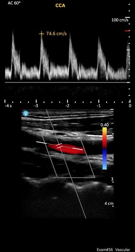
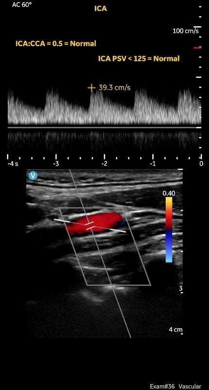
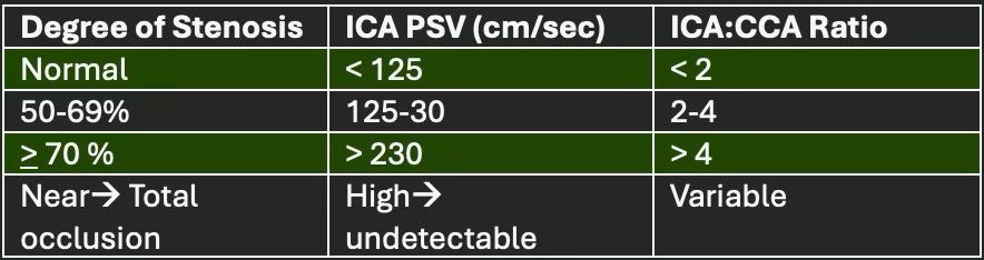
Analyzing carotid stenosis: Using PW Doppler, measure PSV in the CCA and ICA. Analyze PSV in the ICA and calculate ICA: CCA ratio. The most important number to remember is ICA PSV > 125 is abnormal.
#POCUS
#POCUS
December 8, 2024 at 11:14 AM
Analyzing carotid stenosis: Using PW Doppler, measure PSV in the CCA and ICA. Analyze PSV in the ICA and calculate ICA: CCA ratio. The most important number to remember is ICA PSV > 125 is abnormal.
#POCUS
#POCUS