


Joanna Pylvänäinen
@jwpylvanainen.bsky.social
Image analysis | Infrastructure | Education | Post-doctoral researcher
Reposted by Joanna Pylvänäinen

🚀Join Helsinki BioImaging as an Image Analysis Specialist🖥️to support cross-disciplinary image analysis projects. If you love coding and are interested in working at the crossroads of computer science and biology, apply by 🗓️ November 15, 2025⤵️
jobs.helsinki.fi/job/Helsinki...
#jobopening
jobs.helsinki.fi/job/Helsinki...
#jobopening

Image Analysis Specialist/Engineer/Coordinator, Helsinki Bioimaging
Image Analysis Specialist/Engineer/Coordinator, Helsinki Bioimaging
jobs.helsinki.fi
October 10, 2025 at 1:58 PM
🚀Join Helsinki BioImaging as an Image Analysis Specialist🖥️to support cross-disciplinary image analysis projects. If you love coding and are interested in working at the crossroads of computer science and biology, apply by 🗓️ November 15, 2025⤵️
jobs.helsinki.fi/job/Helsinki...
#jobopening
jobs.helsinki.fi/job/Helsinki...
#jobopening
Reposted by Joanna Pylvänäinen
- A postdoc to work on cancer extravasation (4-year project).
- A postdoc help in completing Filopodia-related lab projects (2-year).
- Multiple Marie Curie postdoc positions that will open in the fall.
- We will open a 5-year position to cover my teaching (70% research and 30% teaching).
- A postdoc help in completing Filopodia-related lab projects (2-year).
- Multiple Marie Curie postdoc positions that will open in the fall.
- We will open a 5-year position to cover my teaching (70% research and 30% teaching).
Are you looking for a postdoc?
I will be looking for 2-4 people after the summer to join the lab !!
Do not hesitate to reach out and disseminate !!
I will be looking for 2-4 people after the summer to join the lab !!
Do not hesitate to reach out and disseminate !!
July 1, 2025 at 4:46 AM
- A postdoc to work on cancer extravasation (4-year project).
- A postdoc help in completing Filopodia-related lab projects (2-year).
- Multiple Marie Curie postdoc positions that will open in the fall.
- We will open a 5-year position to cover my teaching (70% research and 30% teaching).
- A postdoc help in completing Filopodia-related lab projects (2-year).
- Multiple Marie Curie postdoc positions that will open in the fall.
- We will open a 5-year position to cover my teaching (70% research and 30% teaching).
Reposted by Joanna Pylvänäinen
Are you looking for a postdoc?
I will be looking for 2-4 people after the summer to join the lab !!
Do not hesitate to reach out and disseminate !!
I will be looking for 2-4 people after the summer to join the lab !!
Do not hesitate to reach out and disseminate !!
June 23, 2025 at 2:29 PM
Are you looking for a postdoc?
I will be looking for 2-4 people after the summer to join the lab !!
Do not hesitate to reach out and disseminate !!
I will be looking for 2-4 people after the summer to join the lab !!
Do not hesitate to reach out and disseminate !!
Reposted by Joanna Pylvänäinen
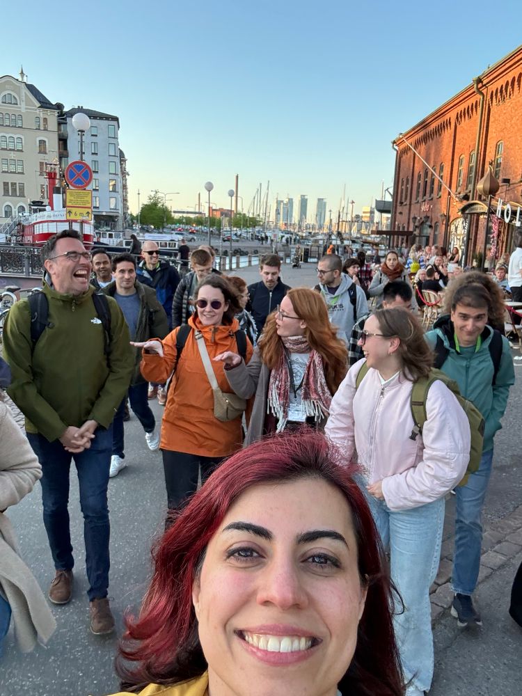
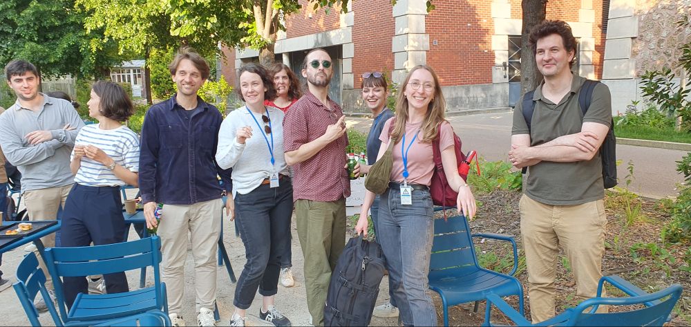
Walking through streets of Helsinki. After-conference social events are the best way to finish the day. Sunny Helsinki summer night, good weather, good company, what else can one ask for? @florianjug.bsky.social @jwpylvanainen.bsky.social @turkubioimaging.bsky.social @ai4life.bsky.social
May 28, 2025 at 8:20 AM
Walking through streets of Helsinki. After-conference social events are the best way to finish the day. Sunny Helsinki summer night, good weather, good company, what else can one ask for? @florianjug.bsky.social @jwpylvanainen.bsky.social @turkubioimaging.bsky.social @ai4life.bsky.social
Reposted by Joanna Pylvänäinen
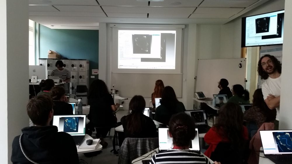
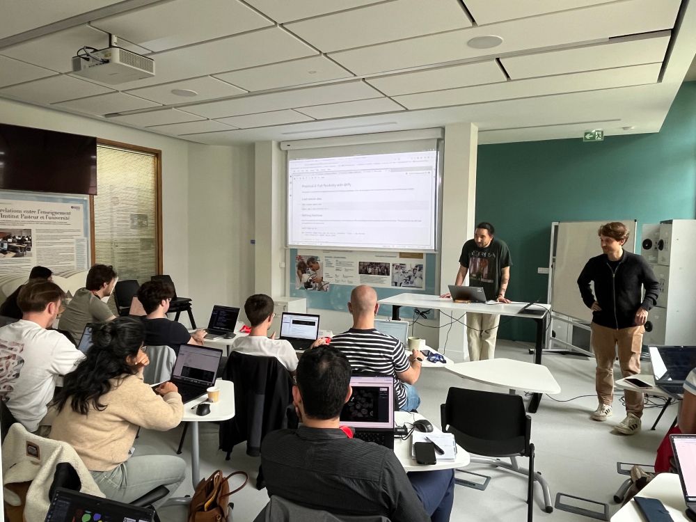
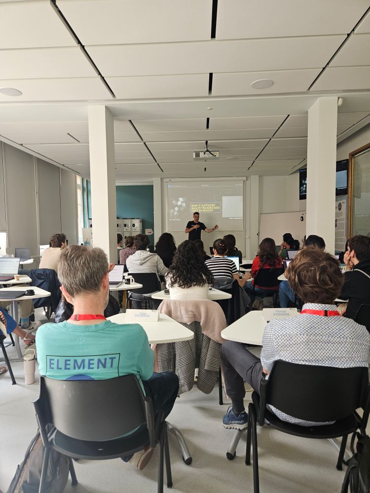
4th day on the go for #neubiaspasteur2025 with a @biapyx.bsky.social practical course by @jwpylvanainen.bsky.social and a #cellpose #dask and RNA2Seg (doi.org/10.1101/2025...) course with @gaellel.bsky.social and Alice Blondel
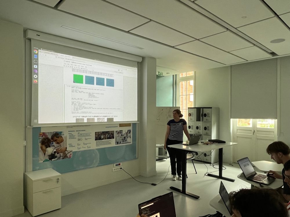
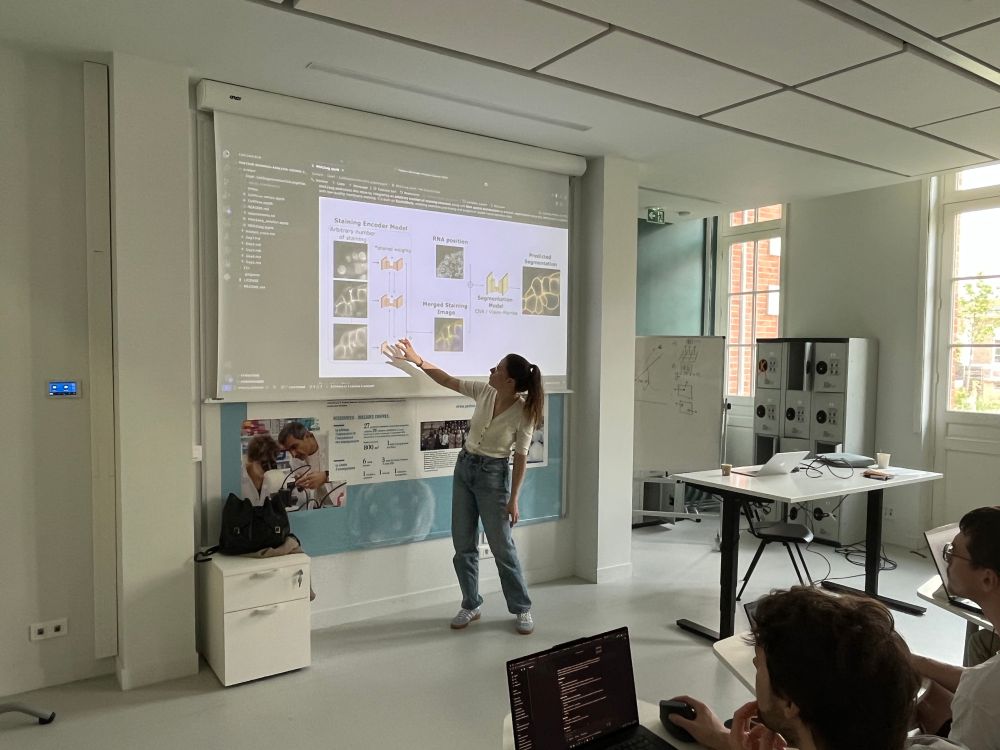
May 15, 2025 at 12:33 PM
4th day on the go for #neubiaspasteur2025 with a @biapyx.bsky.social practical course by @jwpylvanainen.bsky.social and a #cellpose #dask and RNA2Seg (doi.org/10.1101/2025...) course with @gaellel.bsky.social and Alice Blondel
Reposted by Joanna Pylvänäinen
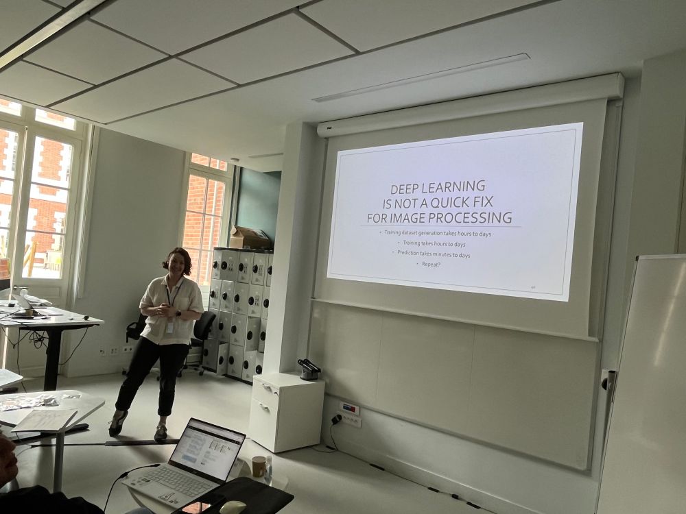
Perfect way to finish the day with ☀️ and warm evening.
Thanks to the teachers and dear colleagues @jwpylvanainen.bsky.social @mambroset.bsky.social @gaellel.bsky.social @m-albert.bsky.social Carlos Garcia-Lopez & Heloise Monnet, and the students that join us !
Thanks to the teachers and dear colleagues @jwpylvanainen.bsky.social @mambroset.bsky.social @gaellel.bsky.social @m-albert.bsky.social Carlos Garcia-Lopez & Heloise Monnet, and the students that join us !
May 15, 2025 at 12:37 PM
Perfect way to finish the day with ☀️ and warm evening.
Thanks to the teachers and dear colleagues @jwpylvanainen.bsky.social @mambroset.bsky.social @gaellel.bsky.social @m-albert.bsky.social Carlos Garcia-Lopez & Heloise Monnet, and the students that join us !
Thanks to the teachers and dear colleagues @jwpylvanainen.bsky.social @mambroset.bsky.social @gaellel.bsky.social @m-albert.bsky.social Carlos Garcia-Lopez & Heloise Monnet, and the students that join us !
Reposted by Joanna Pylvänäinen
Second day with Ilastik (@ilastik-team.bsky.social) covered by @MinhSonPhan, with @strigaud.bsky.social and @jytinevez.bsky.social as helpers. How to easily train a pixel classifier to segment focal adhesions..
May 13, 2025 at 10:21 AM
Second day with Ilastik (@ilastik-team.bsky.social) covered by @MinhSonPhan, with @strigaud.bsky.social and @jytinevez.bsky.social as helpers. How to easily train a pixel classifier to segment focal adhesions..
Reposted by Joanna Pylvänäinen
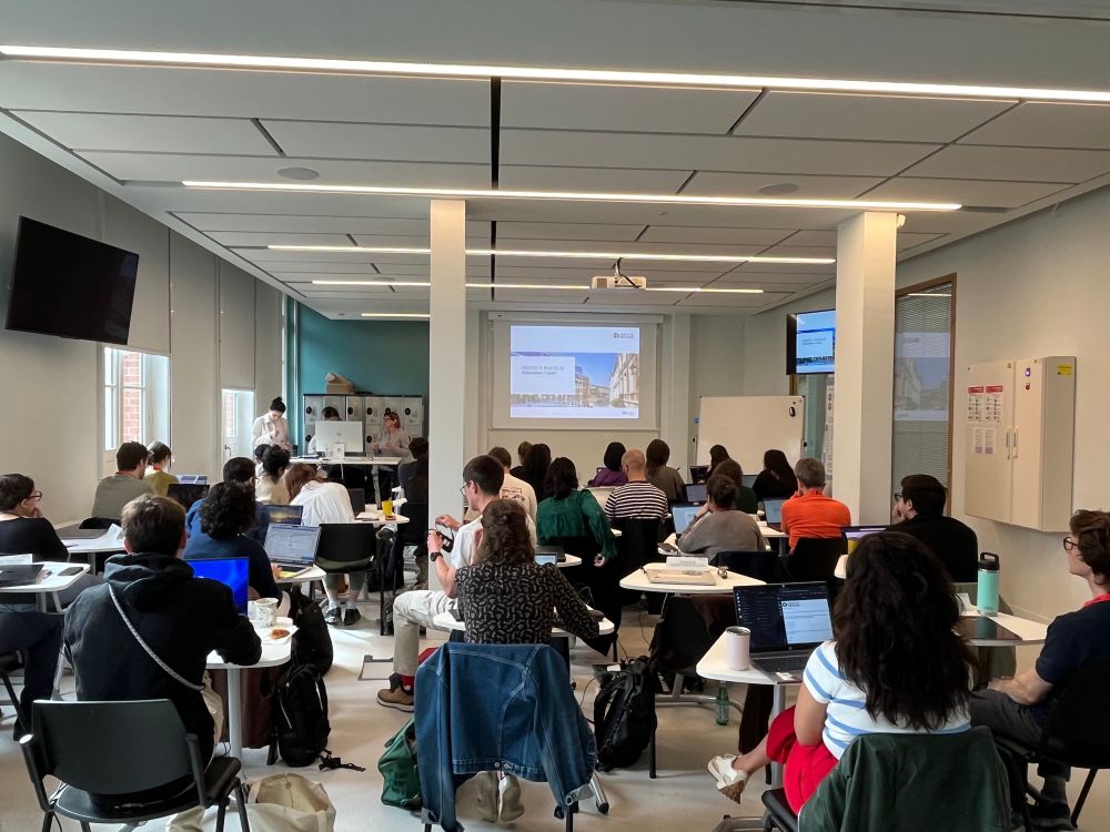
Kick-starting the #neubiaspasteur2025 #BioImageAnalysis courses at @pasteur.fr and @pasteuredu.bsky.social
May 13, 2025 at 7:57 AM
Kick-starting the #neubiaspasteur2025 #BioImageAnalysis courses at @pasteur.fr and @pasteuredu.bsky.social
Just arrived to Institute Pasteur in Paris for Neubias Image analysis course. Todays opening talk about the future of bioimage analysis by @florianjug.bsky.social - very cool week ahead! #neubiaspasteur2025
May 13, 2025 at 7:46 AM
Just arrived to Institute Pasteur in Paris for Neubias Image analysis course. Todays opening talk about the future of bioimage analysis by @florianjug.bsky.social - very cool week ahead! #neubiaspasteur2025
Reposted by Joanna Pylvänäinen
Introducing warpfield, an open source Python library for GPU-accelerated non-rigid 3D registration. Warps and aligns gigavoxel volumes within seconds (not hours). For 3D microscopy, region-to-region and cell-to-cell matching.
A collaboration with @mh123.bsky.social 🚀
github.com/danionella/w...
A collaboration with @mh123.bsky.social 🚀
github.com/danionella/w...
May 12, 2025 at 5:25 AM
Introducing warpfield, an open source Python library for GPU-accelerated non-rigid 3D registration. Warps and aligns gigavoxel volumes within seconds (not hours). For 3D microscopy, region-to-region and cell-to-cell matching.
A collaboration with @mh123.bsky.social 🚀
github.com/danionella/w...
A collaboration with @mh123.bsky.social 🚀
github.com/danionella/w...
Reposted by Joanna Pylvänäinen

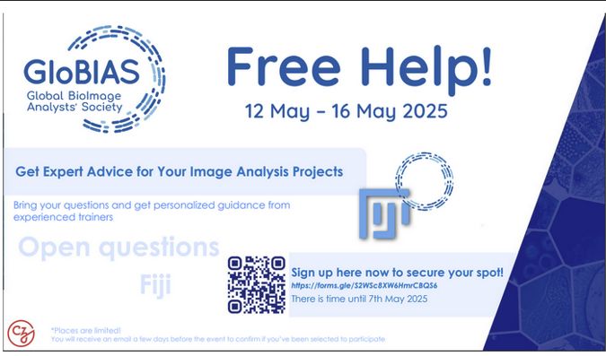
Second round of hashtag#GloBIAS Free Help (previously Call4Help) is happening in May. You can register for the sessions on our webpage: www.globias.org/activities/g...
Registration deadline is 7. May 2025.
Registration deadline is 7. May 2025.
April 28, 2025 at 8:22 AM
Second round of hashtag#GloBIAS Free Help (previously Call4Help) is happening in May. You can register for the sessions on our webpage: www.globias.org/activities/g...
Registration deadline is 7. May 2025.
Registration deadline is 7. May 2025.
Reposted by Joanna Pylvänäinen
If you train people in ImageJ, Fiji, whatever, even if it isn't your day job, please let us know, and please share this post! Thanks a ton, we really appreciate it. (2/2) forms.gle/C8dUpg8qZkbD... #bioimaging #microscopy #bioimageanalysis
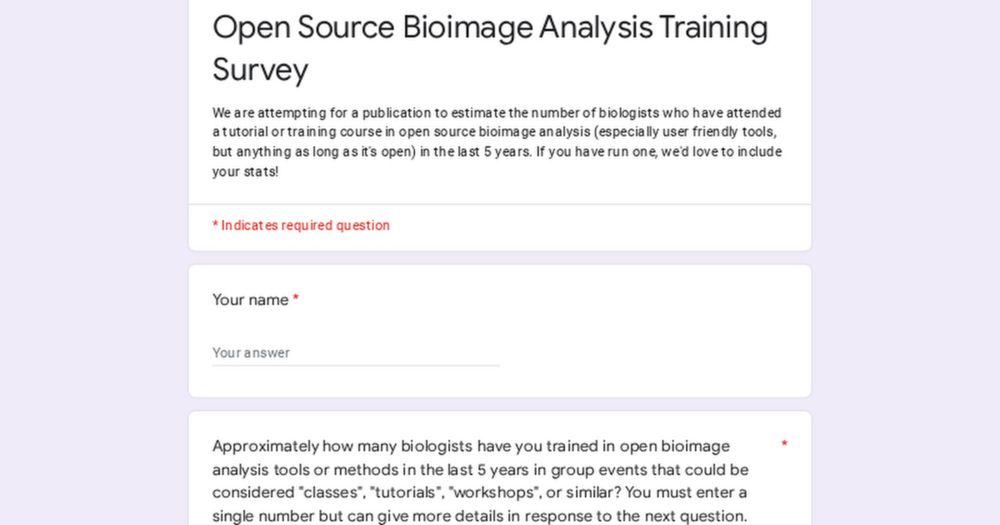
Open Source Bioimage Analysis Training Survey
We are attempting for a publication to estimate the number of biologists who have attended a tutorial or training course in open source bioimage analysis (especially user friendly tools, but anything ...
forms.gle
April 8, 2025 at 2:32 PM
If you train people in ImageJ, Fiji, whatever, even if it isn't your day job, please let us know, and please share this post! Thanks a ton, we really appreciate it. (2/2) forms.gle/C8dUpg8qZkbD... #bioimaging #microscopy #bioimageanalysis
Reposted by Joanna Pylvänäinen
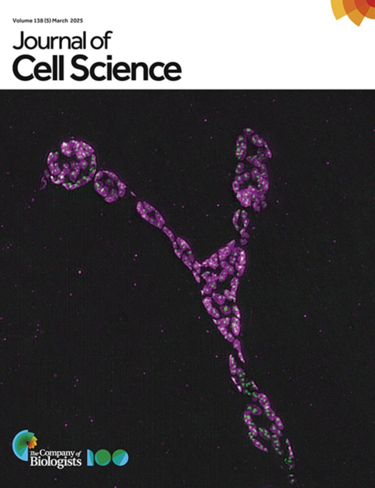
Joanna Pylvänäinen @jwpylvanainen.bsky.social Hanna Grobe and Guillaume Jacquemet @guijacquemet.bsky.social present their practical guidelines for data exploration in quantitative cell biology.
journals.biologists.com/jcs/article/...
#OpenAccess
journals.biologists.com/jcs/article/...
#OpenAccess

April 14, 2025 at 2:01 PM
Joanna Pylvänäinen @jwpylvanainen.bsky.social Hanna Grobe and Guillaume Jacquemet @guijacquemet.bsky.social present their practical guidelines for data exploration in quantitative cell biology.
journals.biologists.com/jcs/article/...
#OpenAccess
journals.biologists.com/jcs/article/...
#OpenAccess
Reposted by Joanna Pylvänäinen
Delighted to announce that @erinweisbart.bsky.social and I have teamed up to create a new bioimage analysis video podcast called Ask Erin/Dear Beth - you can check it out at the link below! It will highlight common challenges in #bioimageanalysis, as well as our favorite solutions to them. (1/x)

Ask Erin, Dear Beth
On Ask Erin/Dear Beth, bioimage analysis experts Beth Cimini and Erin Weisbart, of the Imaging Platform at the Broad Institute of MIT and Harvard, answer your image analysis questions! Whether it’s ab...
www.youtube.com
April 7, 2025 at 10:11 PM
Delighted to announce that @erinweisbart.bsky.social and I have teamed up to create a new bioimage analysis video podcast called Ask Erin/Dear Beth - you can check it out at the link below! It will highlight common challenges in #bioimageanalysis, as well as our favorite solutions to them. (1/x)
We wrote an opinion with simple, practical advice for building data exploration workflows in quantitative cell biology with @guijacquemet.bsky.social and @hanna09.bsky.social . I hope you find it useful!
Here we share our opinions on "Practical considerations for data exploration in quantitative cell biology"
journals.biologists.com/jcs/article/...
Joanna, Hanna and I worked quite a bit on this piece and I hope you find it useful
Many thanks to @jcellsci.bsky.social for publishing it
journals.biologists.com/jcs/article/...
Joanna, Hanna and I worked quite a bit on this piece and I hope you find it useful
Many thanks to @jcellsci.bsky.social for publishing it

Practical considerations for data exploration in quantitative cell biology
Summary: We present practical guidelines for data exploration in quantitative cell biology that are designed to help yield reliable conclusions and promote a collaborative, transparent approach to dat...
journals.biologists.com
April 8, 2025 at 5:26 AM
We wrote an opinion with simple, practical advice for building data exploration workflows in quantitative cell biology with @guijacquemet.bsky.social and @hanna09.bsky.social . I hope you find it useful!
Reposted by Joanna Pylvänäinen
Repost appreciated:
Open #PhDposition in structural virology in my group! Join us in Umeå, 🇸🇪 to uncover how arboviruses remodel the cellular interior. In situ #cryoET combined with #virology, #cellbiology, and #biophysics.
Deadline 4 May. More info: www.carlsonlab.se/join/
Open #PhDposition in structural virology in my group! Join us in Umeå, 🇸🇪 to uncover how arboviruses remodel the cellular interior. In situ #cryoET combined with #virology, #cellbiology, and #biophysics.
Deadline 4 May. More info: www.carlsonlab.se/join/
April 4, 2025 at 8:06 AM
Repost appreciated:
Open #PhDposition in structural virology in my group! Join us in Umeå, 🇸🇪 to uncover how arboviruses remodel the cellular interior. In situ #cryoET combined with #virology, #cellbiology, and #biophysics.
Deadline 4 May. More info: www.carlsonlab.se/join/
Open #PhDposition in structural virology in my group! Join us in Umeå, 🇸🇪 to uncover how arboviruses remodel the cellular interior. In situ #cryoET combined with #virology, #cellbiology, and #biophysics.
Deadline 4 May. More info: www.carlsonlab.se/join/
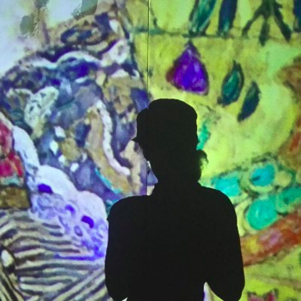
Jamie Hyneman's lecture on creative problem solving & climate innovation was packed, so we found a cozy spot in the streaming room to watch live with @ivanhcenalmor. @aboakademi.bsky.social

April 1, 2025 at 6:58 AM
Jamie Hyneman's lecture on creative problem solving & climate innovation was packed, so we found a cozy spot in the streaming room to watch live with @ivanhcenalmor. @aboakademi.bsky.social
Reposted by Joanna Pylvänäinen
In their Essay, @jwpylvanainen.bsky.social, @guijacquemet.bsky.social and @lankylaste.bsky.social outline their practical recommendation for developing image analysis software for life science applications.
journals.biologists.com/jcs/article/...
journals.biologists.com/jcs/article/...

March 20, 2025 at 10:36 AM
In their Essay, @jwpylvanainen.bsky.social, @guijacquemet.bsky.social and @lankylaste.bsky.social outline their practical recommendation for developing image analysis software for life science applications.
journals.biologists.com/jcs/article/...
journals.biologists.com/jcs/article/...
Reposted by Joanna Pylvänäinen
Excellent set of guidelines for anyone developing software for life scientists, from @jwpylvanainen.bsky.social, @lankylaste.bsky.social and @guijacquemet.bsky.social
journals.biologists.com/jcs/article/...
journals.biologists.com/jcs/article/...
Practical recommendations for developing software for life science applications
ABSTRACT. Developing user-friendly image analysis software is essential for advancing biological and life science research. However, the interdisciplinary gap between software developers and life scie...
journals.biologists.com
March 19, 2025 at 3:56 PM
Excellent set of guidelines for anyone developing software for life scientists, from @jwpylvanainen.bsky.social, @lankylaste.bsky.social and @guijacquemet.bsky.social
journals.biologists.com/jcs/article/...
journals.biologists.com/jcs/article/...
Reposted by Joanna Pylvänäinen
Awesome to have @henriqueslab.bsky.social join us as a visiting professor!!!

We are happy and honored to have Dr. Anna Bigas and Professor Ricardo Henriques as our Visiting Professors. Welcome on board!
#stemcells #cancer #ImagingTechnologies
@utu.fi @turkubioscience.bsky.social @turkubioimaging.bsky.social @henriqueslab.bsky.social @hospitaldelmar.bsky.social
#stemcells #cancer #ImagingTechnologies
@utu.fi @turkubioscience.bsky.social @turkubioimaging.bsky.social @henriqueslab.bsky.social @hospitaldelmar.bsky.social
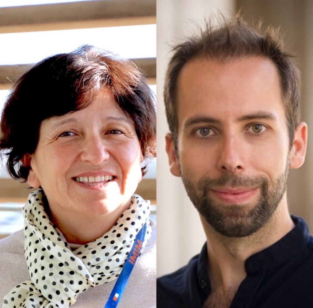
InFLAMES welcomes two new visiting professors - InFLAMES Research Flagship
Dr. Anna Bigas and Professor Ricardo Henriques will join the InFLAMES Flagship as visiting professors at Åbo Akademi University and lend their expertise to strengthen the international InFLAMES network.
inflames.utu.fi
March 17, 2025 at 10:06 AM
Awesome to have @henriqueslab.bsky.social join us as a visiting professor!!!
Reposted by Joanna Pylvänäinen
Check this one out! This is a great read (I am bias).
But Joanna and Stefania did an amazing job putting the article together.
But Joanna and Stefania did an amazing job putting the article together.
Fresh from the press! @lankylaste.bsky.social, @guijacquemet.bsky.social and I put together a practical guide on how to develop software for life science applications.

Practical recommendations for developing software for life science applications
ABSTRACT. Developing user-friendly image analysis software is essential for advancing biological and life science research. However, the interdisciplinary gap between software developers and life scie...
doi.org
March 17, 2025 at 10:08 AM
Check this one out! This is a great read (I am bias).
But Joanna and Stefania did an amazing job putting the article together.
But Joanna and Stefania did an amazing job putting the article together.
Reposted by Joanna Pylvänäinen
And it’s out! Thanks to @bethcimini.bsky.social and all the participants in the @biologists.bsky.social “Effectively Communicating BioImage Analysis” workshop last year for the fruitful discussions that started the idea behind this paper. Get reading and let us know what you think!
Fresh from the press! @lankylaste.bsky.social, @guijacquemet.bsky.social and I put together a practical guide on how to develop software for life science applications.
Practical recommendations for developing software for life science applications
ABSTRACT. Developing user-friendly image analysis software is essential for advancing biological and life science research. However, the interdisciplinary gap between software developers and life scie...
doi.org
March 17, 2025 at 10:16 AM
And it’s out! Thanks to @bethcimini.bsky.social and all the participants in the @biologists.bsky.social “Effectively Communicating BioImage Analysis” workshop last year for the fruitful discussions that started the idea behind this paper. Get reading and let us know what you think!
Fresh from the press! @lankylaste.bsky.social, @guijacquemet.bsky.social and I put together a practical guide on how to develop software for life science applications.
Practical recommendations for developing software for life science applications
ABSTRACT. Developing user-friendly image analysis software is essential for advancing biological and life science research. However, the interdisciplinary gap between software developers and life scie...
doi.org
March 17, 2025 at 9:44 AM
Fresh from the press! @lankylaste.bsky.social, @guijacquemet.bsky.social and I put together a practical guide on how to develop software for life science applications.
Yet another throwback to my thesis work—digging into year-old data. For once, past me did future me a favor: everything nicely documented in one place! 💪🏻

February 5, 2025 at 12:52 PM
Yet another throwback to my thesis work—digging into year-old data. For once, past me did future me a favor: everything nicely documented in one place! 💪🏻