


Katrin Böttcher
@kboettcherlab.bsky.social
physician scientist 👩🏻⚕️🔬
aims at understanding #MAIT cell biology in #tissueimmunity and #cancer
passionate about #immunometabolism
based at M3 Research Center at University of Tübingen
aims at understanding #MAIT cell biology in #tissueimmunity and #cancer
passionate about #immunometabolism
based at M3 Research Center at University of Tübingen
Reposted by Katrin Böttcher
BREAKING: The Nobel Prize in Physiology or Medicine has been awarded jointly to Mary E. Brunkow, Fred Ramsdell, and Shimon Sakaguchi "for their discoveries concerning peripheral immune tolerance"
Stay tuned for more.
#NobelPrize
Stay tuned for more.
#NobelPrize

October 6, 2025 at 9:34 AM
BREAKING: The Nobel Prize in Physiology or Medicine has been awarded jointly to Mary E. Brunkow, Fred Ramsdell, and Shimon Sakaguchi "for their discoveries concerning peripheral immune tolerance"
Stay tuned for more.
#NobelPrize
Stay tuned for more.
#NobelPrize
I am more than happy that our study on MAIT cells in MASLD and their role in liver cancer immunity is now out in Journal of Hepatology!! 🤩
⬇️ Here's a thread about:
🧬 Immunometabolic dysfunction of MAIT cells in MASLD: A novel barrier to liver cancer immunity 🧬
(led by Sebastian Deschler)
⬇️ Here's a thread about:
🧬 Immunometabolic dysfunction of MAIT cells in MASLD: A novel barrier to liver cancer immunity 🧬
(led by Sebastian Deschler)

June 23, 2025 at 7:51 AM
I am more than happy that our study on MAIT cells in MASLD and their role in liver cancer immunity is now out in Journal of Hepatology!! 🤩
⬇️ Here's a thread about:
🧬 Immunometabolic dysfunction of MAIT cells in MASLD: A novel barrier to liver cancer immunity 🧬
(led by Sebastian Deschler)
⬇️ Here's a thread about:
🧬 Immunometabolic dysfunction of MAIT cells in MASLD: A novel barrier to liver cancer immunity 🧬
(led by Sebastian Deschler)
The team is growing - if you’re interested in doing a PhD in translational research at the interface of #immunology, #oncology and #Tcellengineering at the M3 Research Center in Tübingen, follow the link below for more information and apply!
jobs.medizin.uni-tuebingen.de/Job/6357/PhD...
jobs.medizin.uni-tuebingen.de/Job/6357/PhD...
PhD Student in Cancer Immunology
jobs.medizin.uni-tuebingen.de
May 28, 2025 at 9:27 AM
The team is growing - if you’re interested in doing a PhD in translational research at the interface of #immunology, #oncology and #Tcellengineering at the M3 Research Center in Tübingen, follow the link below for more information and apply!
jobs.medizin.uni-tuebingen.de/Job/6357/PhD...
jobs.medizin.uni-tuebingen.de/Job/6357/PhD...
🤩 Thrilled and honoured to talk MAIT cells at the Falk Experimental Days of Hepatology among a fantastic lineup of researchers in Lyon today 🤩#MAITs #hepatology #translation #immunology

April 25, 2025 at 9:57 AM
🤩 Thrilled and honoured to talk MAIT cells at the Falk Experimental Days of Hepatology among a fantastic lineup of researchers in Lyon today 🤩#MAITs #hepatology #translation #immunology
📣 !! JOB ALERT for PhD students !!
My lab is moving to the M3 Research Center at the University of Tübingen and is looking for PhD students who are enthusiastic about translational research.
If you are, read on below ⬇️
My lab is moving to the M3 Research Center at the University of Tübingen and is looking for PhD students who are enthusiastic about translational research.
If you are, read on below ⬇️
March 4, 2025 at 1:33 PM
📣 !! JOB ALERT for PhD students !!
My lab is moving to the M3 Research Center at the University of Tübingen and is looking for PhD students who are enthusiastic about translational research.
If you are, read on below ⬇️
My lab is moving to the M3 Research Center at the University of Tübingen and is looking for PhD students who are enthusiastic about translational research.
If you are, read on below ⬇️
🔬👩🏻⚕️👩🔬🧫⭐️

🌟 Happy Women in Science Day! 👩🔬🔬🧪
Today, we celebrate brilliant women pushing boundaries and driving scientific innovation.
Stay tuned for inspiring stories that celebrate passion, perseverance, and progress. 🚀
#yefis #WomenInScience #immunology
Today, we celebrate brilliant women pushing boundaries and driving scientific innovation.
Stay tuned for inspiring stories that celebrate passion, perseverance, and progress. 🚀
#yefis #WomenInScience #immunology

February 12, 2025 at 10:19 AM
🔬👩🏻⚕️👩🔬🧫⭐️
Reposted by Katrin Böttcher

Don’t forget to apply by 18th Feb for our 3 year Wellcome post-doc to join our great team working on HBV tissue immunity! www.linkedin.com/jobs/view/re...
UCL hiring Research Fellow (Maini Lab) in London, England, United Kingdom | LinkedIn
Posted 10:45:46 AM. About usApplications are invited for a Postdoctoral Research Fellow position in the Division of…See this and similar jobs on LinkedIn.
www.linkedin.com
February 5, 2025 at 4:08 PM
Don’t forget to apply by 18th Feb for our 3 year Wellcome post-doc to join our great team working on HBV tissue immunity! www.linkedin.com/jobs/view/re...
Reposted by Katrin Böttcher
Great new paper from Sandberg group @ki.se in @pnas.org showing IL-10 producing MAIT cells - a population negatively regulated by IL-18
www.pnas.org/doi/10.1073/...
www.pnas.org/doi/10.1073/...
PNAS
Proceedings of the National Academy of Sciences (PNAS), a peer reviewed journal of the National Academy of Sciences (NAS) - an authoritative source of high-impact, original research that broadly spans...
www.pnas.org
February 4, 2025 at 8:43 PM
Great new paper from Sandberg group @ki.se in @pnas.org showing IL-10 producing MAIT cells - a population negatively regulated by IL-18
www.pnas.org/doi/10.1073/...
www.pnas.org/doi/10.1073/...
Reposted by Katrin Böttcher

Looking for an #immunology #postdoc?
Then I *THOROUGHLY RECOMMEND* @mainilab.bsky.social
Incredibly supportive PI & environment
Cutting-edge science🧪 (dissecting hepatic #immunity in people w/ #hepatitisB & #cancer)
Fab team & collabs
Great institute
APPLY NOW: www.linkedin.com/jobs/view/re...
Then I *THOROUGHLY RECOMMEND* @mainilab.bsky.social
Incredibly supportive PI & environment
Cutting-edge science🧪 (dissecting hepatic #immunity in people w/ #hepatitisB & #cancer)
Fab team & collabs
Great institute
APPLY NOW: www.linkedin.com/jobs/view/re...
UCL hiring Research Fellow (Maini Lab) in London, England, United Kingdom | LinkedIn
Posted 10:45:46 AM. About usApplications are invited for a Postdoctoral Research Fellow position in the Division of…See this and similar jobs on LinkedIn.
www.linkedin.com
January 21, 2025 at 1:21 PM
Looking for an #immunology #postdoc?
Then I *THOROUGHLY RECOMMEND* @mainilab.bsky.social
Incredibly supportive PI & environment
Cutting-edge science🧪 (dissecting hepatic #immunity in people w/ #hepatitisB & #cancer)
Fab team & collabs
Great institute
APPLY NOW: www.linkedin.com/jobs/view/re...
Then I *THOROUGHLY RECOMMEND* @mainilab.bsky.social
Incredibly supportive PI & environment
Cutting-edge science🧪 (dissecting hepatic #immunity in people w/ #hepatitisB & #cancer)
Fab team & collabs
Great institute
APPLY NOW: www.linkedin.com/jobs/view/re...
Don’t miss @kakape.bsky.social talking viruses, pandemics and science journalism in Heidelberg if you are around!
Vortrag: Verantwortung und Vertrauen im Wissenschaftsjournalismus – Veranstaltung mit Kai Kupferschmidt, Nature Marsilius Gastprofessor für Wissenschaftskommunikation an der Universität Heidelberg
www.uni-heidelberg.de/de/newsroom/...
www.uni-heidelberg.de/de/newsroom/...

January 14, 2025 at 1:15 PM
Don’t miss @kakape.bsky.social talking viruses, pandemics and science journalism in Heidelberg if you are around!
🌟 joint christmas party with @gmwiedemann-lab.bsky.social and Middelhoff lab was a success - looking forward to keeping the good lab spirit up in 2025 🌟🎄

December 11, 2024 at 9:32 AM
🌟 joint christmas party with @gmwiedemann-lab.bsky.social and Middelhoff lab was a success - looking forward to keeping the good lab spirit up in 2025 🌟🎄
Reposted by Katrin Böttcher
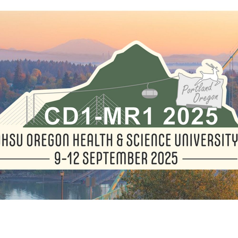
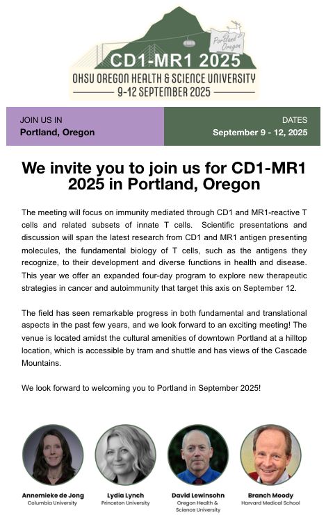
SAVE THE DATES - Join us for CD1-MR1 2025, September 9-12 in Portland, Oregon!
Updates are on the website www.cd1mr1.com including Program at a Glance, Location and Venue details.
Updates are on the website www.cd1mr1.com including Program at a Glance, Location and Venue details.
November 30, 2024 at 7:56 AM
SAVE THE DATES - Join us for CD1-MR1 2025, September 9-12 in Portland, Oregon!
Updates are on the website www.cd1mr1.com including Program at a Glance, Location and Venue details.
Updates are on the website www.cd1mr1.com including Program at a Glance, Location and Venue details.
Reposted by Katrin Böttcher
We’ve jumped on board fully here on 🦋
A starter pack of those working on our favourite cell … the T cell 🥰🙈🥰
If you want an add let us know
go.bsky.app/8B5P25c
A starter pack of those working on our favourite cell … the T cell 🥰🙈🥰
If you want an add let us know
go.bsky.app/8B5P25c
November 19, 2024 at 10:11 PM
We’ve jumped on board fully here on 🦋
A starter pack of those working on our favourite cell … the T cell 🥰🙈🥰
If you want an add let us know
go.bsky.app/8B5P25c
A starter pack of those working on our favourite cell … the T cell 🥰🙈🥰
If you want an add let us know
go.bsky.app/8B5P25c
Reposted by Katrin Böttcher

ICYMI: earlier this year in #Science #Immunology, Josh Gray, Donna Farber & al. analyzed the phenotype, transcriptome, function, & repertoire of #gdTcells in mucosal & lymphoid tissues & blood from 176 #human donors across the life span! Subjects ranged in age from just a few days to over 80 years!

Human γδ T cells in diverse tissues exhibit site-specific maturation dynamics across the life span
Human tissue γδ T cells are diverse and functionally heterogeneous in early life and differentiate into disseminated effectors in adults.
scim.ag
November 27, 2024 at 9:38 PM
ICYMI: earlier this year in #Science #Immunology, Josh Gray, Donna Farber & al. analyzed the phenotype, transcriptome, function, & repertoire of #gdTcells in mucosal & lymphoid tissues & blood from 176 #human donors across the life span! Subjects ranged in age from just a few days to over 80 years!