


@obcbioimaging.bsky.social
Finally,congratulations to Carmen Mata Mangas for winning the poster prize

January 7, 2026 at 5:47 PM
Finally,congratulations to Carmen Mata Mangas for winning the poster prize
The final talk of the day: Mark Johnston of Syngenta on using microscopy in crop protection research

January 7, 2026 at 4:31 PM
The final talk of the day: Mark Johnston of Syngenta on using microscopy in crop protection research
Sam Caddell from MOA Technology explaining how the GALAXY platform aids novel herbicide discovery and understanding how these new herbicides work

January 7, 2026 at 4:15 PM
Sam Caddell from MOA Technology explaining how the GALAXY platform aids novel herbicide discovery and understanding how these new herbicides work
Nikon's Aki Suto demonstrating the @picoquant.bsky.social FLIM system on our AX NSPARC confocal

January 7, 2026 at 3:49 PM
Nikon's Aki Suto demonstrating the @picoquant.bsky.social FLIM system on our AX NSPARC confocal
Flash talks! 📸

January 7, 2026 at 3:35 PM
Flash talks! 📸
Mark Fricker now speaking about quantitative organelle imaging in plants

January 7, 2026 at 2:46 PM
Mark Fricker now speaking about quantitative organelle imaging in plants
Secondly, Petra Boevink from The James Hutton Institute on plant pathogens, secreted RXLR effectors and the pitfalls of imaging infected plants

January 7, 2026 at 2:33 PM
Secondly, Petra Boevink from The James Hutton Institute on plant pathogens, secreted RXLR effectors and the pitfalls of imaging infected plants
Time for the short talks- first, Joanna Chustecki on imaging the dynamics of plant mitochondria

January 7, 2026 at 2:09 PM
Time for the short talks- first, Joanna Chustecki on imaging the dynamics of plant mitochondria

Post-lunch, @ajcellbio.bsky.social telling us about imaging plant endocytosis using multiple techniques 🔬

January 7, 2026 at 1:17 PM
Post-lunch, @ajcellbio.bsky.social telling us about imaging plant endocytosis using multiple techniques 🔬
AX NSPARC demos underway!

January 7, 2026 at 12:41 PM
AX NSPARC demos underway!
Now speaking is Ho-Wei Wu of @slcuplants.bsky.social about imaging of circadian gene expression in live growing root tips


January 7, 2026 at 11:51 AM
Now speaking is Ho-Wei Wu of @slcuplants.bsky.social about imaging of circadian gene expression in live growing root tips
@obcbioimaging.bsky.social 's very own Charlotte Pain telling us about imaging the very dynamic plant endoplasmic reticulum

January 7, 2026 at 11:07 AM
@obcbioimaging.bsky.social 's very own Charlotte Pain telling us about imaging the very dynamic plant endoplasmic reticulum
Next up Ray Wightman of @slcuplants.bsky.social talking about confocal, FLIM and Raman microscopy of plants🌿🔬
January 7, 2026 at 10:46 AM
Next up Ray Wightman of @slcuplants.bsky.social talking about confocal, FLIM and Raman microscopy of plants🌿🔬
Thank you to @obu-hst-research.bsky.social Pro Vice-Chancellor Prof Astrid Schloerscheidt for opening the conference
January 7, 2026 at 10:35 AM
Thank you to @obu-hst-research.bsky.social Pro Vice-Chancellor Prof Astrid Schloerscheidt for opening the conference
First talk at Flora in Focus- Gail Preston @gmpreston.bsky.social of University of Oxford, speaking about metal hyperaccumulation and plant pathogen protection

January 7, 2026 at 10:30 AM
First talk at Flora in Focus- Gail Preston @gmpreston.bsky.social of University of Oxford, speaking about metal hyperaccumulation and plant pathogen protection
Not just flurries of snow in Oxford- flurries of activity as we set up for Flora in Focus: A Microscopy Conference for Plant Scientists


January 5, 2026 at 2:28 PM
Not just flurries of snow in Oxford- flurries of activity as we set up for Flora in Focus: A Microscopy Conference for Plant Scientists
Christmas- the time of year you get out the fancy stuff 🔬💎
(The History of Science Museum in Oxford has some nice blingy things)
(The History of Science Museum in Oxford has some nice blingy things)

December 19, 2025 at 11:24 AM
Christmas- the time of year you get out the fancy stuff 🔬💎
(The History of Science Museum in Oxford has some nice blingy things)
(The History of Science Museum in Oxford has some nice blingy things)
Reposted

It's certainly been a busy year at the RMS - and you can read more about what we've been up to over the last 12 months in our special 'Annual Highlights' feature in the latest issue of infocus Magazine #RMSinfocus 🙌
Read more: https://ow.ly/ogpE50XImkl
Read more: https://ow.ly/ogpE50XImkl

December 12, 2025 at 11:46 AM
It's certainly been a busy year at the RMS - and you can read more about what we've been up to over the last 12 months in our special 'Annual Highlights' feature in the latest issue of infocus Magazine #RMSinfocus 🙌
Read more: https://ow.ly/ogpE50XImkl
Read more: https://ow.ly/ogpE50XImkl
We’re excited to announce our special guest speaker for Flora in Focus: A Microscopy Conference for Plant Scientists
Join us as Dr Alex Johnson, Research Fellow at University of Exeter, presents “Probing plant endocytosis at multiple scales.”
Join us as Dr Alex Johnson, Research Fellow at University of Exeter, presents “Probing plant endocytosis at multiple scales.”

December 5, 2025 at 10:11 AM
We’re excited to announce our special guest speaker for Flora in Focus: A Microscopy Conference for Plant Scientists
Join us as Dr Alex Johnson, Research Fellow at University of Exeter, presents “Probing plant endocytosis at multiple scales.”
Join us as Dr Alex Johnson, Research Fellow at University of Exeter, presents “Probing plant endocytosis at multiple scales.”
Reposted
🔬 THREAD: UK Bioimaging User Access Fund is OPEN!
Up to £5k for imaging experiments, £2k for image analysis
Access world-class facilities at Crick, ESRIC, KCL, Liverpool, Oxford Brookes & York
First deadline: 9 Dec 2025 📅
🧵👇
Up to £5k for imaging experiments, £2k for image analysis
Access world-class facilities at Crick, ESRIC, KCL, Liverpool, Oxford Brookes & York
First deadline: 9 Dec 2025 📅
🧵👇

October 24, 2025 at 1:27 PM
🔬 THREAD: UK Bioimaging User Access Fund is OPEN!
Up to £5k for imaging experiments, £2k for image analysis
Access world-class facilities at Crick, ESRIC, KCL, Liverpool, Oxford Brookes & York
First deadline: 9 Dec 2025 📅
🧵👇
Up to £5k for imaging experiments, £2k for image analysis
Access world-class facilities at Crick, ESRIC, KCL, Liverpool, Oxford Brookes & York
First deadline: 9 Dec 2025 📅
🧵👇
We are pleased to announce the full list of speakers for Flora in Focus: A Microscopy Conference for Plant Scientists.
Sign up now to reserve your spot! bit.ly/42fqd3y
Sign up now to reserve your spot! bit.ly/42fqd3y

December 2, 2025 at 12:55 PM
We are pleased to announce the full list of speakers for Flora in Focus: A Microscopy Conference for Plant Scientists.
Sign up now to reserve your spot! bit.ly/42fqd3y
Sign up now to reserve your spot! bit.ly/42fqd3y
🎤 Meet our speaker: Sam Caddell!
As a scientist at MOA Technology, he will be sharing "GALAXY a high content imaging platform enabling novel herbicide discovery " - a session you won’t want to miss!
As a scientist at MOA Technology, he will be sharing "GALAXY a high content imaging platform enabling novel herbicide discovery " - a session you won’t want to miss!

November 27, 2025 at 10:49 AM
🎤 Meet our speaker: Sam Caddell!
As a scientist at MOA Technology, he will be sharing "GALAXY a high content imaging platform enabling novel herbicide discovery " - a session you won’t want to miss!
As a scientist at MOA Technology, he will be sharing "GALAXY a high content imaging platform enabling novel herbicide discovery " - a session you won’t want to miss!
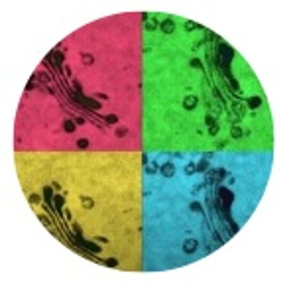
With submissions invited for the 2026 BioImaging Awards, we thought we would share some of the 'comedy' images submitted in previous years #bioimaging #oxfordbrookes #graduateresearchsymposium

November 26, 2025 at 1:06 PM
With submissions invited for the 2026 BioImaging Awards, we thought we would share some of the 'comedy' images submitted in previous years #bioimaging #oxfordbrookes #graduateresearchsymposium
Reposted
Registration is *NOW OPEN* for the 21st International Microscopy Congress, taking place in Liverpool from 31 August - 4 September 2026 🙌 🔬 🙂
Book now: https://www.imc21.org.uk/
Book now: https://www.imc21.org.uk/

November 24, 2025 at 3:44 PM
Registration is *NOW OPEN* for the 21st International Microscopy Congress, taking place in Liverpool from 31 August - 4 September 2026 🙌 🔬 🙂
Book now: https://www.imc21.org.uk/
Book now: https://www.imc21.org.uk/