


Kevin Keel
@ucpath.bsky.social
Veterinary pathologist, conservation advocate, wildlife-disease scientist, outdoorsman. And I love parasites, but especially the metazoans!
Scientific, one-health and naturalist content.
Scientific, one-health and naturalist content.
Reposted by Kevin Keel
A Super Ager, Fauja Singh, ran his 1st marathon at age 89, and 8 more after that through age 101.
How does he die at age 114 years?
Hit by a car.
apnews.com/article/olde...
How does he die at age 114 years?
Hit by a car.
apnews.com/article/olde...

July 16, 2025 at 9:44 PM
A Super Ager, Fauja Singh, ran his 1st marathon at age 89, and 8 more after that through age 101.
How does he die at age 114 years?
Hit by a car.
apnews.com/article/olde...
How does he die at age 114 years?
Hit by a car.
apnews.com/article/olde...
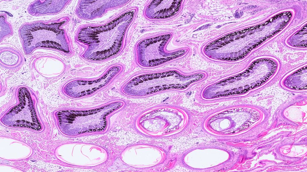
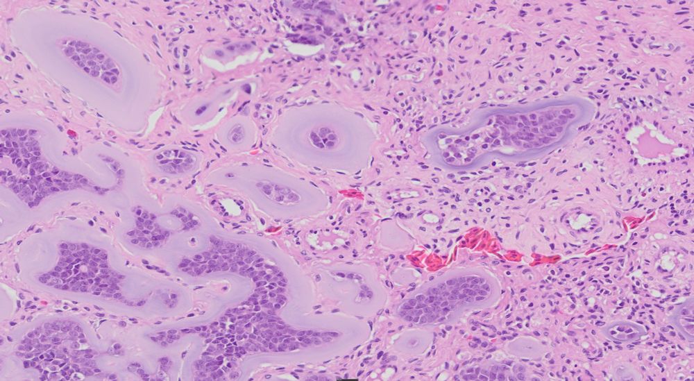
Beautiful representation of feather follicles from a chickadee. A happy accident from the histology lab. They are not supposed to be sectioned obliquely but it makes them look pretty interesting in an abstract sort of way. 🪶
July 16, 2025 at 9:42 PM
Beautiful representation of feather follicles from a chickadee. A happy accident from the histology lab. They are not supposed to be sectioned obliquely but it makes them look pretty interesting in an abstract sort of way. 🪶
Submit your case for the Wildlife Pathology Workshop at #WDA2025
📅 Deadline for slide submission: June 9
🔬 Focus: Gross & histopathology of wildlife cases
📍 Held during the WDA Annual Meeting
#VeterinaryPathology #WildlifeHealth #OneHealth
wda2025.com/event/wda-an...
📅 Deadline for slide submission: June 9
🔬 Focus: Gross & histopathology of wildlife cases
📍 Held during the WDA Annual Meeting
#VeterinaryPathology #WildlifeHealth #OneHealth
wda2025.com/event/wda-an...
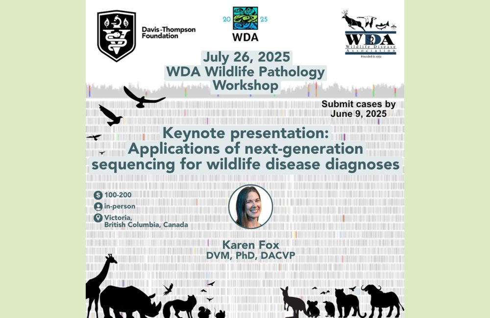
WDA and Davis-Thompson Foundation, Annual Wildlife Pathology Workshop - Wildlife Disease Association 2025
The event begins with a keynote lecture by Dr. Karen Fox, entitled “Applications of next-generation sequencing for wildlife disease diagnoses.” The rest of the day will be a histopathology workshop. P...
wda2025.com
May 9, 2025 at 4:20 PM
Submit your case for the Wildlife Pathology Workshop at #WDA2025
📅 Deadline for slide submission: June 9
🔬 Focus: Gross & histopathology of wildlife cases
📍 Held during the WDA Annual Meeting
#VeterinaryPathology #WildlifeHealth #OneHealth
wda2025.com/event/wda-an...
📅 Deadline for slide submission: June 9
🔬 Focus: Gross & histopathology of wildlife cases
📍 Held during the WDA Annual Meeting
#VeterinaryPathology #WildlifeHealth #OneHealth
wda2025.com/event/wda-an...
With 1,000 dairy herds affected by avian influenza and the disease regularly changing, the absence of this program is going to hurt.
www.reuters.com/business/hea...
www.reuters.com/business/hea...
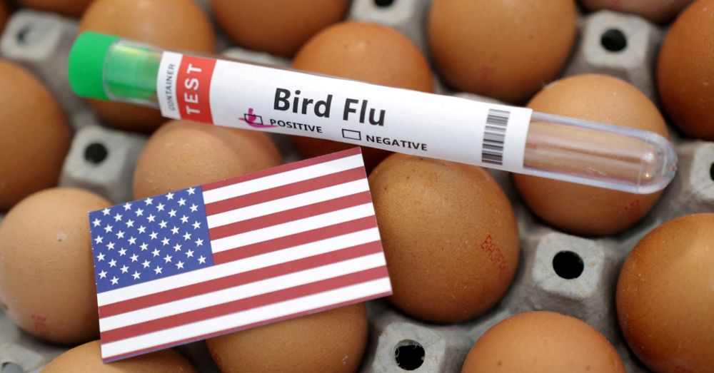
FDA suspends program to improve bird flu testing due to staff cuts
The U.S. Food and Drug Administration is suspending efforts to improve its bird flu testing of milk, cheese and pet food due to massive staff cuts at the agency, according to an email seen by Reuters and a source familiar with the situation.
www.reuters.com
April 3, 2025 at 11:45 PM
With 1,000 dairy herds affected by avian influenza and the disease regularly changing, the absence of this program is going to hurt.
www.reuters.com/business/hea...
www.reuters.com/business/hea...
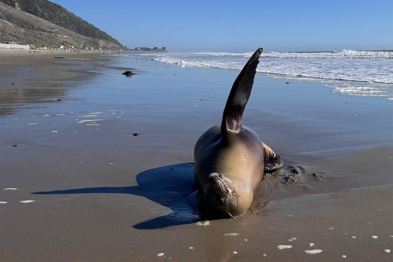
California is no stranger to domoic acid, but this year it's much worse much earlier.
Stranded pinnipeds often exhibit listlessness, head bobbing, disorientation and seizures. Mortality rates are estimated to be ~25%
www.fisheries.noaa.gov/feature-stor...
#VetMed #MarineMammal #VetPath
Stranded pinnipeds often exhibit listlessness, head bobbing, disorientation and seizures. Mortality rates are estimated to be ~25%
www.fisheries.noaa.gov/feature-stor...
#VetMed #MarineMammal #VetPath
April 1, 2025 at 6:28 PM
California is no stranger to domoic acid, but this year it's much worse much earlier.
Stranded pinnipeds often exhibit listlessness, head bobbing, disorientation and seizures. Mortality rates are estimated to be ~25%
www.fisheries.noaa.gov/feature-stor...
#VetMed #MarineMammal #VetPath
Stranded pinnipeds often exhibit listlessness, head bobbing, disorientation and seizures. Mortality rates are estimated to be ~25%
www.fisheries.noaa.gov/feature-stor...
#VetMed #MarineMammal #VetPath
Well, shit.
This is going to cost us a lot more than the money saved. For the price of 38 Abrams tanks, #FAO has gotten a hell of a lot done with support from the U.S.
kbhbradio.com/funding-term...
#VetMed #AnimalHealth #OneHealth
This is going to cost us a lot more than the money saved. For the price of 38 Abrams tanks, #FAO has gotten a hell of a lot done with support from the U.S.
kbhbradio.com/funding-term...
#VetMed #AnimalHealth #OneHealth
Funding terminated for preparedness, response to animal disease outbreaks
WASHINGTON, D.C. - The U.S government had terminated funding for programs at the United Nations Food and Agriculture Organization, (FAO) a spokesperson said. “FAO has received termination notices for ...
kbhbradio.com
March 31, 2025 at 2:37 PM
Well, shit.
This is going to cost us a lot more than the money saved. For the price of 38 Abrams tanks, #FAO has gotten a hell of a lot done with support from the U.S.
kbhbradio.com/funding-term...
#VetMed #AnimalHealth #OneHealth
This is going to cost us a lot more than the money saved. For the price of 38 Abrams tanks, #FAO has gotten a hell of a lot done with support from the U.S.
kbhbradio.com/funding-term...
#VetMed #AnimalHealth #OneHealth
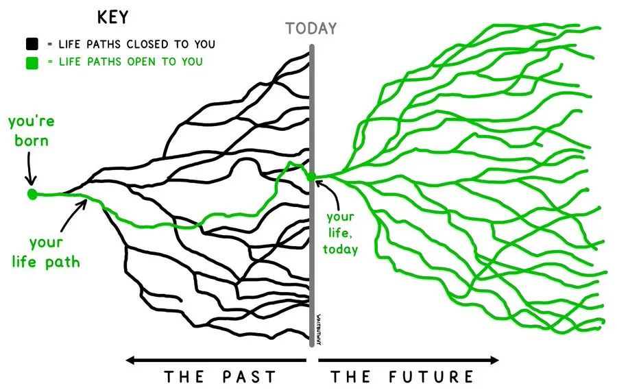
This seems a bit obvious, but it's a very cool illustration of the idea. I tried to figure out where it was first published, but all I found were many social media posts with the image. Guilty!
The future is a web of branching paths, and each choice opens up new possibilities as it closes others. This is as true for a civilization as it is for an individual. The more our collective intelligence can inform each choice, the better our chances of finding paths through to a beautiful future.
March 31, 2025 at 2:25 PM
This seems a bit obvious, but it's a very cool illustration of the idea. I tried to figure out where it was first published, but all I found were many social media posts with the image. Guilty!
Reposted by Kevin Keel

The future is a web of branching paths, and each choice opens up new possibilities as it closes others. This is as true for a civilization as it is for an individual. The more our collective intelligence can inform each choice, the better our chances of finding paths through to a beautiful future.
March 31, 2025 at 6:22 AM
The future is a web of branching paths, and each choice opens up new possibilities as it closes others. This is as true for a civilization as it is for an individual. The more our collective intelligence can inform each choice, the better our chances of finding paths through to a beautiful future.
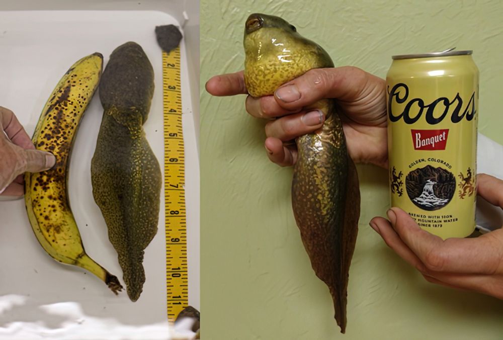
Not a new article, but it's worth posting again just in case you missed it. Bullfrogs are normally tadpoles for 2 or 3 years. Something disrupted this one's metamorphosis so it just kept on growing! I love the references for scale!
#frogs #wildlife
www.americanscientist.org/blog/from-th...
#frogs #wildlife
www.americanscientist.org/blog/from-th...
March 25, 2025 at 7:30 PM
Not a new article, but it's worth posting again just in case you missed it. Bullfrogs are normally tadpoles for 2 or 3 years. Something disrupted this one's metamorphosis so it just kept on growing! I love the references for scale!
#frogs #wildlife
www.americanscientist.org/blog/from-th...
#frogs #wildlife
www.americanscientist.org/blog/from-th...
If only we had a vaccine for ignorance. But, wait! We do!!
Or, at least we did. Unfortunately, critical thinking and education, it would seem, are passé.
www.nbcnews.com/health/healt...
Or, at least we did. Unfortunately, critical thinking and education, it would seem, are passé.
www.nbcnews.com/health/healt...
How the anti-vaccine movement weaponized a 6-year-old's measles death
Anti-vaccine influencers see the child’s death as proof — not of the danger of measles, but of their own debunked theories.
www.nbcnews.com
March 20, 2025 at 7:15 PM
If only we had a vaccine for ignorance. But, wait! We do!!
Or, at least we did. Unfortunately, critical thinking and education, it would seem, are passé.
www.nbcnews.com/health/healt...
Or, at least we did. Unfortunately, critical thinking and education, it would seem, are passé.
www.nbcnews.com/health/healt...
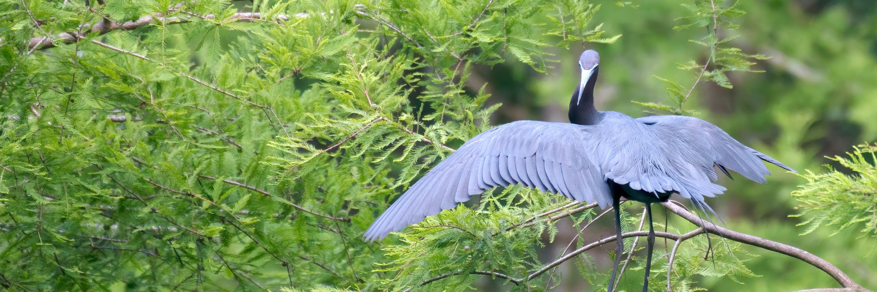
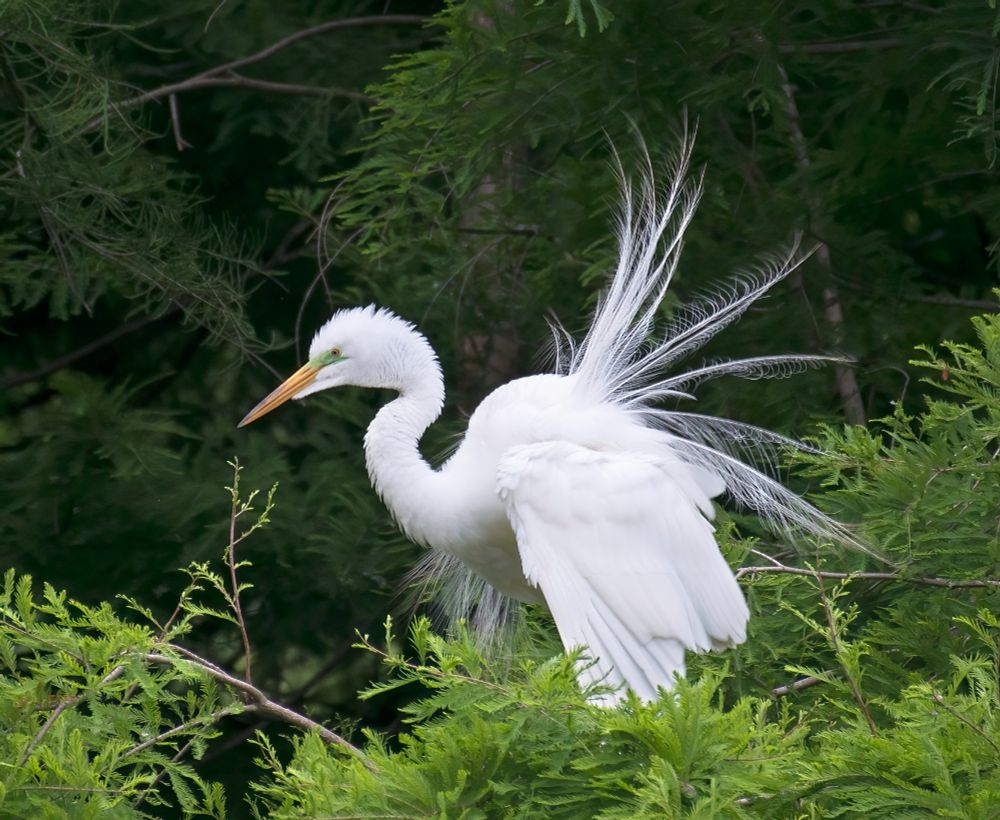
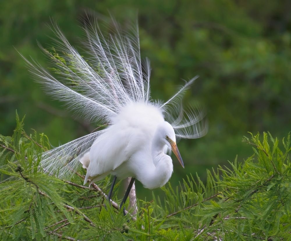
An exhibitionist egret on display. These courtship threads beat millinery any day. Populations of common #egrets (and other spp.) were once decimated so we could put their nuptial plumes in women’s hats. Goshen Swamp, Liberty Co, Georgia.
#birds #wildlife #NaturePhotography #Photography
#birds #wildlife #NaturePhotography #Photography
March 20, 2025 at 5:55 PM
An exhibitionist egret on display. These courtship threads beat millinery any day. Populations of common #egrets (and other spp.) were once decimated so we could put their nuptial plumes in women’s hats. Goshen Swamp, Liberty Co, Georgia.
#birds #wildlife #NaturePhotography #Photography
#birds #wildlife #NaturePhotography #Photography
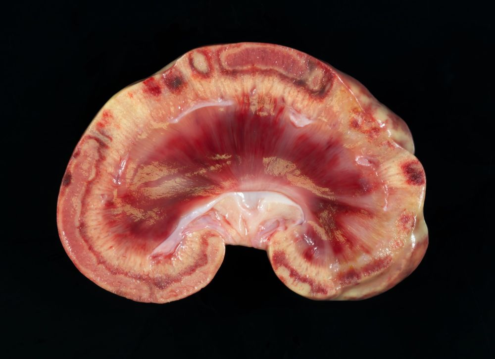
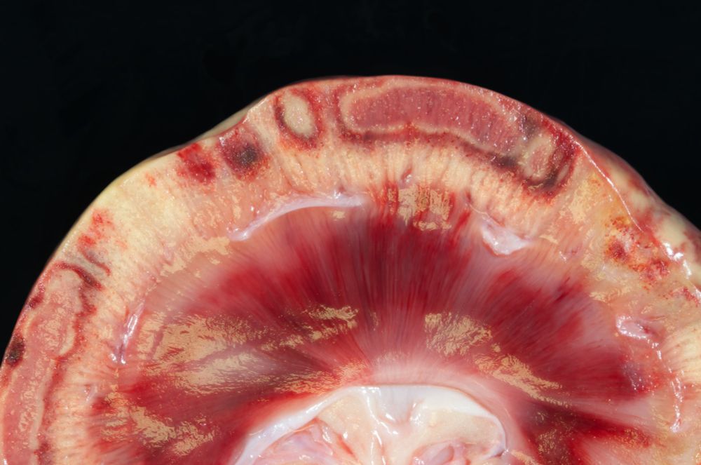
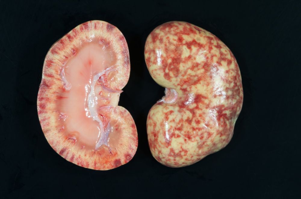
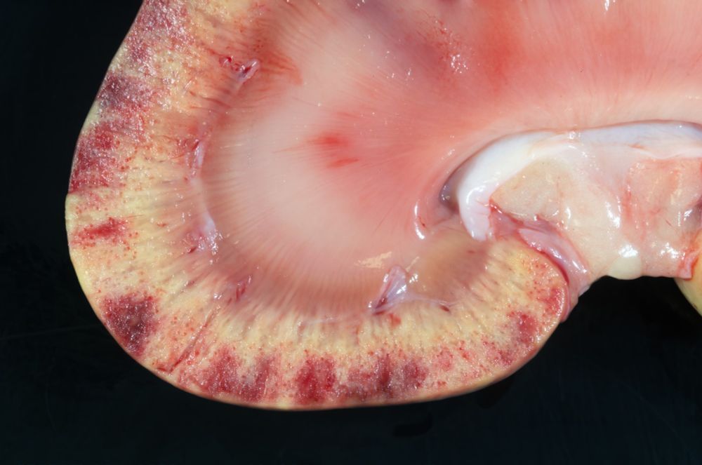
These are some crazy kidneys! You don't have to be a pathologist to think these lesions are beautiful!
I have suspicions as to pathogenesis, but histo will tell.
9-yo dog. Clinicians susp granulomatous osteomyelitis of lumbar v but no gross lesions were evident.
#pathsky #vetpath #RenalPath
I have suspicions as to pathogenesis, but histo will tell.
9-yo dog. Clinicians susp granulomatous osteomyelitis of lumbar v but no gross lesions were evident.
#pathsky #vetpath #RenalPath
March 19, 2025 at 11:29 PM
These are some crazy kidneys! You don't have to be a pathologist to think these lesions are beautiful!
I have suspicions as to pathogenesis, but histo will tell.
9-yo dog. Clinicians susp granulomatous osteomyelitis of lumbar v but no gross lesions were evident.
#pathsky #vetpath #RenalPath
I have suspicions as to pathogenesis, but histo will tell.
9-yo dog. Clinicians susp granulomatous osteomyelitis of lumbar v but no gross lesions were evident.
#pathsky #vetpath #RenalPath
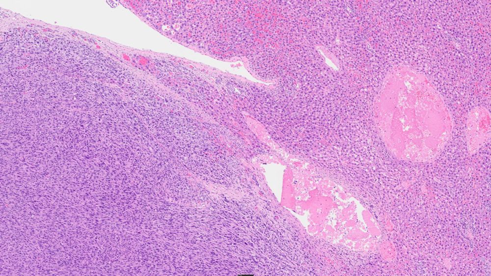
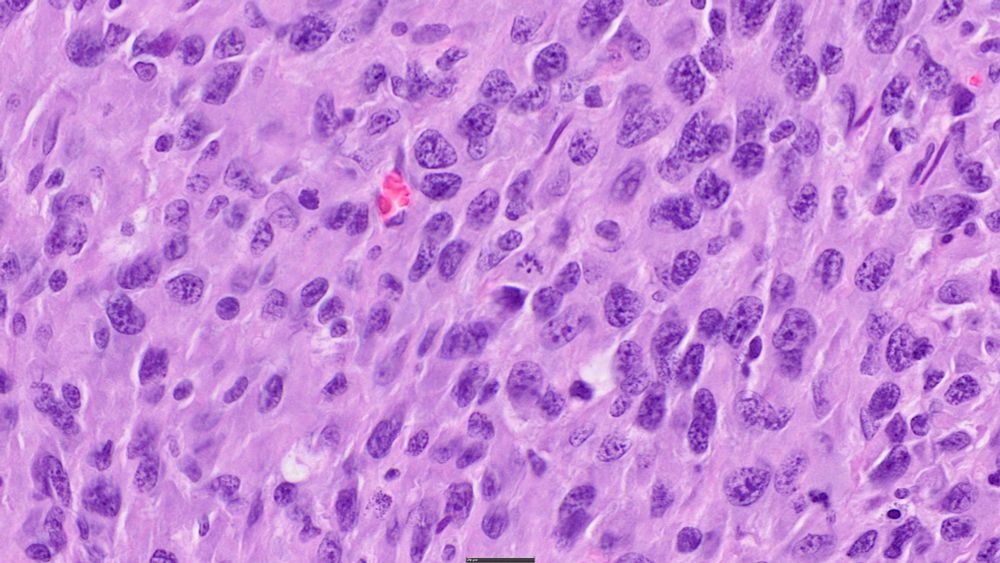
Here's a case you don’t see every day! A hepatic sarcoma in a rat caused by tapeworm larvae. Taenia taeniaformis infestation with chronic inflammation and spindle-cell proliferation paves the way for neoplastic transformation.
Nature is weird sometimes!
#skypath #vetpath #parasites
Nature is weird sometimes!
#skypath #vetpath #parasites
March 18, 2025 at 11:51 PM
Here's a case you don’t see every day! A hepatic sarcoma in a rat caused by tapeworm larvae. Taenia taeniaformis infestation with chronic inflammation and spindle-cell proliferation paves the way for neoplastic transformation.
Nature is weird sometimes!
#skypath #vetpath #parasites
Nature is weird sometimes!
#skypath #vetpath #parasites
I should have mentioned that RFK is the other well known cat who had a tapeworm larvae in his brain.
This cat will survive. Luckily, he's not likely to seek a career in politics. That was a pretty big hole in his brain!
#VetPath #Histopathology #Parasites
bsky.app/profile/ucpa...
This cat will survive. Luckily, he's not likely to seek a career in politics. That was a pretty big hole in his brain!
#VetPath #Histopathology #Parasites
bsky.app/profile/ucpa...
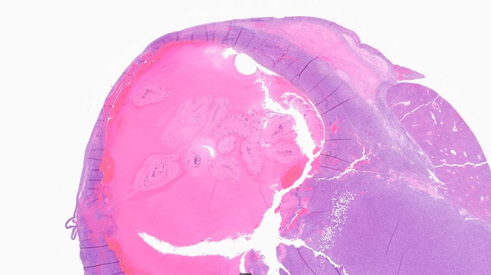
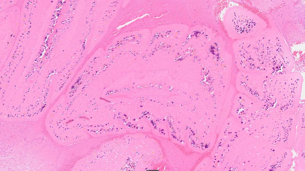
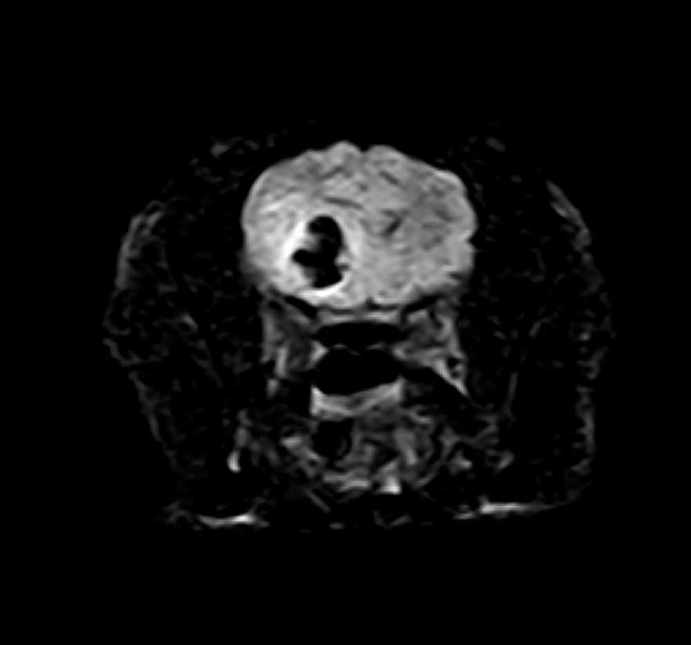
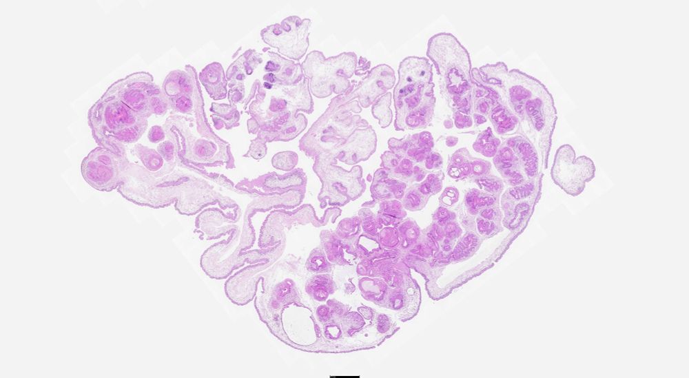
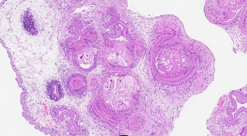
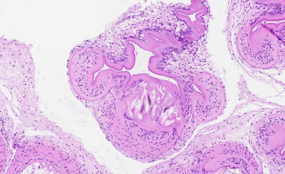
Coolest biopsy of the week! A coenurus from the brain of a cat. This tapeworm larvae has multiple invaginated scolices.
The cat was doing well when discharged 7 days post op.
#VetPath #PathSky #Parasites #Histopathology
The cat was doing well when discharged 7 days post op.
#VetPath #PathSky #Parasites #Histopathology
March 14, 2025 at 3:26 PM
I should have mentioned that RFK is the other well known cat who had a tapeworm larvae in his brain.
This cat will survive. Luckily, he's not likely to seek a career in politics. That was a pretty big hole in his brain!
#VetPath #Histopathology #Parasites
bsky.app/profile/ucpa...
This cat will survive. Luckily, he's not likely to seek a career in politics. That was a pretty big hole in his brain!
#VetPath #Histopathology #Parasites
bsky.app/profile/ucpa...
Coolest biopsy of the week! A coenurus from the brain of a cat. This tapeworm larvae has multiple invaginated scolices.
The cat was doing well when discharged 7 days post op.
#VetPath #PathSky #Parasites #Histopathology
The cat was doing well when discharged 7 days post op.
#VetPath #PathSky #Parasites #Histopathology
March 8, 2025 at 12:25 AM
Coolest biopsy of the week! A coenurus from the brain of a cat. This tapeworm larvae has multiple invaginated scolices.
The cat was doing well when discharged 7 days post op.
#VetPath #PathSky #Parasites #Histopathology
The cat was doing well when discharged 7 days post op.
#VetPath #PathSky #Parasites #Histopathology
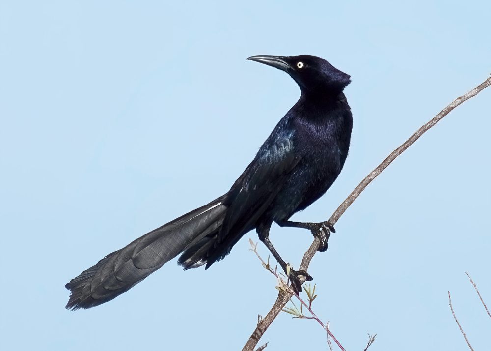
The great-tailed grackle. My most favorite of all the grackles. Look at that tail! Look at that beak! He is seems pretty proud of the whole package. Woodland, CA.
I wonder how long before they try to change the name to Quiscalus americanus.
#birds #wildlifephotography #naturephotography
I wonder how long before they try to change the name to Quiscalus americanus.
#birds #wildlifephotography #naturephotography
February 28, 2025 at 6:09 AM
The great-tailed grackle. My most favorite of all the grackles. Look at that tail! Look at that beak! He is seems pretty proud of the whole package. Woodland, CA.
I wonder how long before they try to change the name to Quiscalus americanus.
#birds #wildlifephotography #naturephotography
I wonder how long before they try to change the name to Quiscalus americanus.
#birds #wildlifephotography #naturephotography
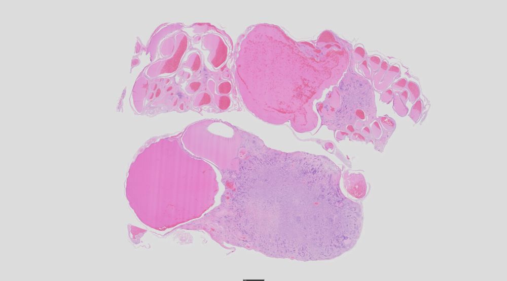
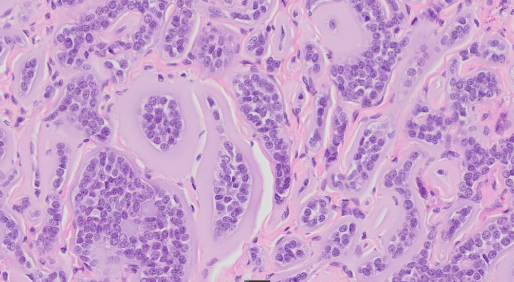
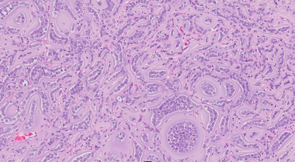
Looking for opinions on this (alleged) ovarian mass from a garter snake. I presume the amphophilic material is redundant basement membrane. Could this be a granulosa cell tumor? We have seen those in snakes (incl garter snakes) but this one is odd.
#pathsky #vetpath #veterinary #tumor #repro
#pathsky #vetpath #veterinary #tumor #repro
February 27, 2025 at 11:59 PM
Looking for opinions on this (alleged) ovarian mass from a garter snake. I presume the amphophilic material is redundant basement membrane. Could this be a granulosa cell tumor? We have seen those in snakes (incl garter snakes) but this one is odd.
#pathsky #vetpath #veterinary #tumor #repro
#pathsky #vetpath #veterinary #tumor #repro
What's your diagnosis? American crow with a lesion on the back of the shank. I've seen this bird every day for a week and a half.
#VetPath #AvianDisease #Veterinary #DermPath
#VetPath #AvianDisease #Veterinary #DermPath
February 18, 2025 at 10:22 PM
What's your diagnosis? American crow with a lesion on the back of the shank. I've seen this bird every day for a week and a half.
#VetPath #AvianDisease #Veterinary #DermPath
#VetPath #AvianDisease #Veterinary #DermPath
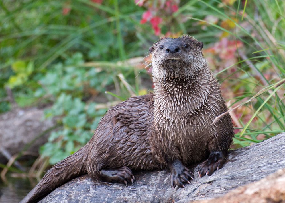
Normally we go to Lake Solano (Yolo Co, California) to see the migrator waterfowl, especially the buffleheads and goldeneye. This day the #RiverOtters were putting on more of a show.
#mammals #WildlifePhotography #Nikon #wildlife
#mammals #WildlifePhotography #Nikon #wildlife
February 5, 2025 at 2:04 AM
Normally we go to Lake Solano (Yolo Co, California) to see the migrator waterfowl, especially the buffleheads and goldeneye. This day the #RiverOtters were putting on more of a show.
#mammals #WildlifePhotography #Nikon #wildlife
#mammals #WildlifePhotography #Nikon #wildlife
A new kind of box #fish? Nope! It's much more interesting. This fishy cell has been stained with an antibody specific for rabies. They don't call it neurotropic for nothing! Antigen fills the cytoplasm but spares the nucleus creating a stark contrast betw the two.🧪
#histopath #PathSky #VetPath
#histopath #PathSky #VetPath

January 31, 2025 at 12:10 AM
A new kind of box #fish? Nope! It's much more interesting. This fishy cell has been stained with an antibody specific for rabies. They don't call it neurotropic for nothing! Antigen fills the cytoplasm but spares the nucleus creating a stark contrast betw the two.🧪
#histopath #PathSky #VetPath
#histopath #PathSky #VetPath
It's a couple of years old but this is a great review of #rabies #virus #pathogenesis and #immune evasion. A student's question on antibody response prompted me to dig a little deeper in the literature. Who doesn't appreciate a good review article?
onlinelibrary.wiley.com/doi/full/10....
onlinelibrary.wiley.com/doi/full/10....

Neuroimmunology of rabies: New insights into an ancient disease
Rabies is an ancient neuroinvasive viral (genus Lyssavirus, family Rhabdoviridae) disease affecting approximately 59,000 people worldwide. The central nervous system (CNS) is targeted, and rabies has...
onlinelibrary.wiley.com
January 30, 2025 at 11:50 PM
It's a couple of years old but this is a great review of #rabies #virus #pathogenesis and #immune evasion. A student's question on antibody response prompted me to dig a little deeper in the literature. Who doesn't appreciate a good review article?
onlinelibrary.wiley.com/doi/full/10....
onlinelibrary.wiley.com/doi/full/10....
Reposted by Kevin Keel

It's "What Is Your Diagnosis?" Wednesday!
What are your differential diagnoses for a flock of 20,000 Bovans Brown laying hens that experienced a 2-week period of increasing mortality starting at 54 weeks of age? Full case: doi.org/10.2460/javm...
What are your differential diagnoses for a flock of 20,000 Bovans Brown laying hens that experienced a 2-week period of increasing mortality starting at 54 weeks of age? Full case: doi.org/10.2460/javm...

January 29, 2025 at 9:56 PM
It's "What Is Your Diagnosis?" Wednesday!
What are your differential diagnoses for a flock of 20,000 Bovans Brown laying hens that experienced a 2-week period of increasing mortality starting at 54 weeks of age? Full case: doi.org/10.2460/javm...
What are your differential diagnoses for a flock of 20,000 Bovans Brown laying hens that experienced a 2-week period of increasing mortality starting at 54 weeks of age? Full case: doi.org/10.2460/javm...
A beautiful cleared example of Fascioloides magna that was submitted to the vet school. The large liver fluke leads to condemnation of beef livers and can cause significant disease in sheep and goats. I see it in white-tailed #deer, who don't seem affected.
#veterinary #parasitology #trematode
#veterinary #parasitology #trematode

January 27, 2025 at 8:52 PM
A beautiful cleared example of Fascioloides magna that was submitted to the vet school. The large liver fluke leads to condemnation of beef livers and can cause significant disease in sheep and goats. I see it in white-tailed #deer, who don't seem affected.
#veterinary #parasitology #trematode
#veterinary #parasitology #trematode
Here is an awesome case from our #histopathology conference this morning!
Lung from a #macaque. What's your dx? The residents hate it when I say, "It's a classic". But it is! 😊
#VetPath #PathSky #lung #parasites
Lung from a #macaque. What's your dx? The residents hate it when I say, "It's a classic". But it is! 😊
#VetPath #PathSky #lung #parasites


January 24, 2025 at 7:03 PM
Here is an awesome case from our #histopathology conference this morning!
Lung from a #macaque. What's your dx? The residents hate it when I say, "It's a classic". But it is! 😊
#VetPath #PathSky #lung #parasites
Lung from a #macaque. What's your dx? The residents hate it when I say, "It's a classic". But it is! 😊
#VetPath #PathSky #lung #parasites