Adrien Heymans
@adrienheymans.bsky.social
200 followers 320 following 24 posts
Postdoc @UPSC on plant anatomy
Posts Media Videos Starter Packs
Pinned
adrienheymans.bsky.social
🌱🧩 How do plant cells get their shape? 🧪

Check out the new #open-access #review exploring the mechanochemical duet between auxin & the cell wall in shaping diverse plant cell types from @srobertgroup.bsky.social's Lab.

🔗 doi.org/10.1111/ppl....

#PlantBiology #CellShape #Auxin #Biomechanics
Epidermal cells acquire different cell shapes to enable their functions and maintain tissue integrity in plants. Meristematic cells differentiate and expand into diverse mature cell types, including jigsaw puzzle-shaped pavement cells (a), round stomatal guard cells (b), elongated epidermal cells in hypocotyls and the apical hook (c), and root epidermal cells with root hairs (d). The spatial distribution and arrangement of cell wall polysaccharides, such as cellulose microfibrils, xyloglucans, and pectins (e.g., homogalacturonan represent as HG), and the abundance of methylester groups on these components in differentiating cells regulate anisotropy during cell growth, enabling the acquisition of specific cell shapes. In addition, proteins like KATANIN and CLASP reorient dynamically the cortical microtubules. These cortical microtubules rearrangement is in response to mechanical cues, either self-generated (as in b, c) or from neighboring cells (a, c), leading to the resulting cell shape.

Attribution-NonCommercial-NoDerivatives 4.0 International

Auxin controls cell expansion by regulating cell wall biosynthesis and cell remodeling. Auxin promotes cell expansion by acidifying the cell wall and activating wall synthesis and loosening enzymes. Auxin efflux (PINs) and influx (AUX1) transporters establish concentration gradients in growing tissue. Auxin enters cells via influx transporters and activates the TRANSPORT INHIBITOR RESPONSE 1/AUXIN SIGNALING F-BOX PROTEINS-AUXIN/INDOLE ACETIC ACID (TIR1/AFB-Aux/IAA) nuclear signaling cascade, which regulates auxin-responsive genes, including AUXIN RESPONSE FACTORs (ARFs) and SMALL AUXIN UP RNAs (SAURs). Auxin activates the H+-ATPase proton pump through TRANSMEMBRANE KINASE 1 (TMK1), acidifying the cell wall and triggering loosening enzymes including PECTIN METHYLESTERASEs (PMEs), EXPANSINs, and XYLOGLUCAN:XYLOGLUCOSYL TRANSFERASEs (EXTs). Cellulose microfibrils are synthesized by the plasma membrane-bound cellulose synthase complex (CSC), with cortical microtubules guiding the exocytosis of this complex toward expanding cell edges. Auxin coordinates the reorientation of cortical microtubules and actin filaments to regulate the trafficking of cell wall polysaccharides to ensure proper cell wall expansion and specific cell shape acquisition.

Attribution-NonCommercial-NoDerivatives 4.0 International
Reposted by Adrien Heymans
umeaplantsciencecentre.se
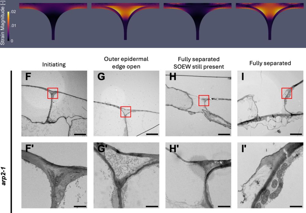
🧪🌾News - How do seedlings straighten when emerging from the soil?🔬

@srobertgroup.bsky.social & collaborators have identified a protein that promotes this seedling straightening via the plant hormone auxin.

Read more about the story published in @pnas.org here👇:
www.upsc.se/about-upsc/n...
Eleven pale seedlings grown next to each other in a row with their yellowish tip bend downwards. Photo: Qian Ma
adrienheymans.bsky.social
🌱🧩 How do plant cells get their shape? 🧪

Check out the new #open-access #review exploring the mechanochemical duet between auxin & the cell wall in shaping diverse plant cell types from @srobertgroup.bsky.social's Lab.

🔗 doi.org/10.1111/ppl....

#PlantBiology #CellShape #Auxin #Biomechanics
Epidermal cells acquire different cell shapes to enable their functions and maintain tissue integrity in plants. Meristematic cells differentiate and expand into diverse mature cell types, including jigsaw puzzle-shaped pavement cells (a), round stomatal guard cells (b), elongated epidermal cells in hypocotyls and the apical hook (c), and root epidermal cells with root hairs (d). The spatial distribution and arrangement of cell wall polysaccharides, such as cellulose microfibrils, xyloglucans, and pectins (e.g., homogalacturonan represent as HG), and the abundance of methylester groups on these components in differentiating cells regulate anisotropy during cell growth, enabling the acquisition of specific cell shapes. In addition, proteins like KATANIN and CLASP reorient dynamically the cortical microtubules. These cortical microtubules rearrangement is in response to mechanical cues, either self-generated (as in b, c) or from neighboring cells (a, c), leading to the resulting cell shape.

Attribution-NonCommercial-NoDerivatives 4.0 International

Auxin controls cell expansion by regulating cell wall biosynthesis and cell remodeling. Auxin promotes cell expansion by acidifying the cell wall and activating wall synthesis and loosening enzymes. Auxin efflux (PINs) and influx (AUX1) transporters establish concentration gradients in growing tissue. Auxin enters cells via influx transporters and activates the TRANSPORT INHIBITOR RESPONSE 1/AUXIN SIGNALING F-BOX PROTEINS-AUXIN/INDOLE ACETIC ACID (TIR1/AFB-Aux/IAA) nuclear signaling cascade, which regulates auxin-responsive genes, including AUXIN RESPONSE FACTORs (ARFs) and SMALL AUXIN UP RNAs (SAURs). Auxin activates the H+-ATPase proton pump through TRANSMEMBRANE KINASE 1 (TMK1), acidifying the cell wall and triggering loosening enzymes including PECTIN METHYLESTERASEs (PMEs), EXPANSINs, and XYLOGLUCAN:XYLOGLUCOSYL TRANSFERASEs (EXTs). Cellulose microfibrils are synthesized by the plasma membrane-bound cellulose synthase complex (CSC), with cortical microtubules guiding the exocytosis of this complex toward expanding cell edges. Auxin coordinates the reorientation of cortical microtubules and actin filaments to regulate the trafficking of cell wall polysaccharides to ensure proper cell wall expansion and specific cell shape acquisition.

Attribution-NonCommercial-NoDerivatives 4.0 International
adrienheymans.bsky.social
🧪🔓 New article from @mutezahmed.bsky.social and @hannahschneider.bsky.social Lab:

"𝐍𝐨𝐝𝐞 𝐨𝐟 𝐨𝐫𝐢𝐠𝐢𝐧 𝐦𝐚𝐭𝐭𝐞𝐫𝐬"⁣
Tina Koehler et al. show that drought responses in maize root anatomy vary by nodal origin.

🔗 doi.org/10.1093/aob/...

#Maize #RootBiology #PlantScience 🌱
Node of origin matters: comparative analysis of soil water limitation effects on nodal root anatomy in maize (Zea mays L.)

Cropped Fig. 7 Slope of the GLM (Fig. 2) characterizing the change in a root anatomical phene across nodes (from CR1-CR5) between drought treatments per genotype as integrative drought response indicator. Transparent colors indicate that a phene did not significantly change across nodes (i.e., with age , Fig. 2, GLM p-value > 0.05). Significance levels of treatment differences are given for each root anatomical phene per genotype (p<0.1. , 0.05*, 0.001**, 0.0001***, not significant (>0.1), ns).

Tina Koehler, Yunhee Kim, Shu-Yin Tung, Adrien Heymans, Nicolas Tyborski, Franziska Steiner, Andreas J Wild, Johanna Pausch, Mutez A Ahmed, Hannah M Schneider, Node of origin matters: comparative analysis of soil water limitation effects on nodal root anatomy in maize (Zea mays L.), Annals of Botany, 2025;, mcaf075, https://doi.org/10.1093/aob/mcaf075
Reposted by Adrien Heymans
aribidopsis.bsky.social
🍀🔬

Recent preprint from Chris Ambrose's lab!

The Arabidopsis WAVE/SCAR Protein BRICK1 Associates with Cell Edges and Plasmodesmata @biorxiv-plants.bsky.social

www.biorxiv.org/content/10.1...
Reposted by Adrien Heymans
guillaumelobet.bsky.social
🧪🌱💡 #PlantSciJob

Are you passionate about #robust cropping systems? Do you want to explore how #plant #roots impact water flow across different scales?

I am thrilled to say that we have 4 open positions (2 #PhD, 2 #postdocs) @UCLouvain (BE) for the EU project DROOGHT.

More: drooght.github.io
Illustration featuring a series of icons representing different concepts: a camera, a bar graph with mathematical symbols, a wheat stalk, a computer monitor, a shovel, a plant growing in a field, interlocking gears, and an Open Science symbol
adrienheymans.bsky.social
🧫Does anyone know of database for 2D plant anatomical data?

I’m especially thinking of datasets from: CellSeT, Icy (.xml) or .zip ImageJ ROIs (e.g., via Celer, PaCeQuant, Cellpose, etc.)

Something like what’s being done with ggPlantMap would be amazing!

#PlantScience #OpenScience
2D plant anatomy data

Pavement cells from Arabidopsis
Root cross section (Pascut et al., 2021: https://doi.org/10.1038/s41467-021-24913-z)
root tip (https://www.future-science.com/doi/10.2144/000114621)
Embryo (https://doi.org/10.1111/nph.12267)
Lateral root emergence (https://doi.org/10.3389/fpls.2019.00206)
Inflorescence stem cross section (https://academic.oup.com/plcell/article/33/2/200/6017181)

ggplantmap @leonardojo.bsky.social (Leonardo and Kajala, 2024: https://doi.org/10.1093/jxb/erae043)
Reposted by Adrien Heymans
guillaumelobet.bsky.social
The 2nd edition of 𝐈𝐧𝐭𝐞𝐫𝐧𝐚𝐭𝐢𝐨𝐧𝐚𝐥 𝐒𝐮𝐦𝐦𝐞𝐫 𝐒𝐜𝐡𝐨𝐨𝐥 𝐨𝐧 𝐌𝐨𝐝𝐞𝐥𝐢𝐧𝐠 𝐖𝐚𝐭𝐞𝐫 𝐅𝐥𝐮𝐱𝐞𝐬 𝐢𝐧 𝐭𝐡𝐞 𝐒𝐨𝐢𝐥-𝐏𝐥𝐚𝐧𝐭 𝐒𝐲𝐬𝐭𝐞𝐦 will be held at UCLouvain, BE, 8-12 Sept 2025.

The course will focus on water flow in the soil-rhizosphere-plant system, plant physiology and rhizosphere hydraulics.

www.uclouvain.be/en/research-...
banner for the summer school with the name and date on it
Reposted by Adrien Heymans
aobp.bsky.social
🌿 New article in @aobp.bsky.social
Authors examined the bark of Euphorbia tirucalli across developmental stages and discovered a unique mode of bark dilatation, allowing prolonged stem photosynthesis.

Full #openaccess
👉 doi.org/pf42

#PlantScience
The image illustrates the arrangement of stomata located within deep stomatal grooves on the juvenile bark of leafless twig shoots of Euphorbia tirucalli. The authors captured this photomicrograph using scanning electron microscopy (SEM, TESCAN, VegaTS software).
adrienheymans.bsky.social
New preprint from @stephanevrg.bsky.social 's lab!

🧪 Simulations + mutant tests show a shift in how we think about plant cell adhesion, challenging "textbook" view on the role of middle lamella.

📄 doi.org/10.1101/2025...

Thank you Özer Erguvan and all co-authors 🔬

#PlantSci #CellBiology
FEM and TEM reveal the process by which cell separation may initiate in the outer epidermal edge filling (OEEF) and propagate to the supracellular outer epidermal wall FEM and TEM reveal the process by which cell separation may initiate in the outer epidermal edge filling (OEEF) and propagate to the supracellular outer epidermal wall (SOEW).
CC-BY-NC-ND 4.0 International license.
(which was not certified by peer review) is the author/funder, who has granted bioRxiv a license to display the preprint in perpetuity. It is
bioRxiv preprint doi: https://doi.org/10.1101/2025.04.03.646819; this version posted April 6, 2025.
Reposted by Adrien Heymans
cellwalldynamics.com
And continuing with conference news—postdoc Bastien Dauphin just gave a great talk at the UK Plant Biomechanics Conference 2025 🎤🌿

He’s using chemo-optogenetics to trigger local changes in the plant cell wall. New tools, new insights!

#PlantBiomechanics #CellWallDynamics #Optogenetics
Bastien Dauphin during his presentation at the UK Plant Biomechanics Conference 2025. Thanks Yoselin Benitez Alfonso for the picture :)
adrienheymans.bsky.social
2nd Summer School on Advanced Soil Physics: Soil-Plant-Water Flow 🧪🌱💧
@agrosphere-ibg-3.bsky.social
@UCLouvain - ELI

Designed for master’s and PhD students, as well as early-career scientists in fields such as soil physics, plant physiology, water management, and environmental science

#ISMC
https://soil-modeling.org/activities/summer-school/summer-school-advanced-soil-physics
adrienheymans.bsky.social
🌟 In bonus, there is some FEM with #BVPy of Single cells with fragmented or fused vacuole.

Thanks @srobertgroup.bsky.social for introducing me to this strange world 🔬

Check out the Jupyter Notebook 🖥️ and try it for yourself:
github.com/SRobertGroup...
FEM analysis of a single biological cell with a fragmented vacuole. Color gradient depicts the strain magnitude.
The cell geometry is a square with rounded corners, where:
Length = 6 µm; Corner radius = 1 µm; Cell wall thickness= 2 µm.
adrienheymans.bsky.social
🧪🌱Preprint: "Regulation of vacuole fusion, a pivotal mechanism mitigating salt-induced inhibition of root cell growth"

It shows that low-salt stress inhibits vacuole fusion, but may serve as a protective mechanism!

📜Betz et al., 2025 dx.doi.org/10.21203/rs....

#PlantSciences
Fig. 1: Vacuoles respond to salt stress. Confocal images of root cells from Col-0 YFP-VAMP711 treated with 0 mM or 75 mM NaCl  for 24 h, showing YFP fluorescence. (b’-b’’) Magnification of (Fig1.a), highlighting vacuole convolution in the medial plane. (c-c "”) 3D projections of YFP-VAMP711 at 0 mM (c") or 75 mM NaCl (c""), showing how a full reconstruction can mask vacuole convolution.
adrienheymans.bsky.social
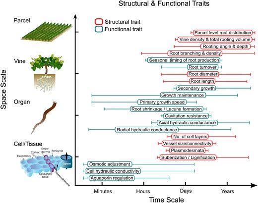
REVIEW: "Root system ideotypes: what is the potential for breeding drought-tolerant grapevine rootstocks?" 🍇

They summarized nicely the #FSPM challenges of mixing different scales of time and space.

📜 Bernardo et al., 2025 doi.org/10.1093/jxb/...
@jxbotany.bsky.social

#PlantSciences 🧪
Root structural and functional traits that interact to bring about root system behavior under drought. One reason root system behavior is so challenging to study and predict is that it results from a complex interaction of structural (red) and functional (cyan) traits that interact across different scales of time (x-axis) and space (y-axis). Even single traits can manifest across different scales of time (whiskers). For example, a complex functional trait like radial hydraulic conductance can change over very short time frames (i.e. minutes to hours) via changes in aquaporin regulation and cell hydraulic conductivity, and over very long time frames (i.e. days to years) via changes in suberization, cell number, and the extent of secondary growth.
Reposted by Adrien Heymans
adrienheymans.bsky.social
Sankey plot.
For #R users there is a great and easy #ggplot library {ggsankey} to make these kind of graphs:
github.com/davidsjoberg...

#DataViz
Reposted by Adrien Heymans
effiebastounis.bsky.social
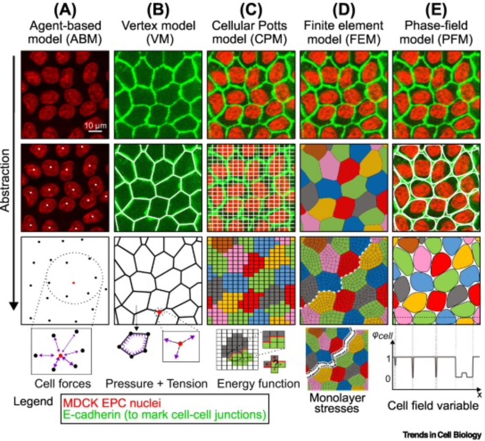
Delighted that our review on modeling of #endothelial and #epithelial #cell monolayer #biomechanics is now published in @cp-trendscellbio.bsky.social: doi.org/10.1016/j.tc...! Thanks to all contributors and to DFG, Universität Tübingen, @cmfi.bsky.social for the support!
Reposted by Adrien Heymans
aafkegros.bsky.social
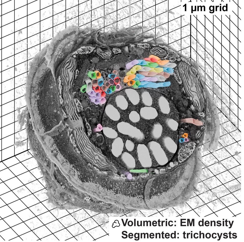
Microscopy Nodes is now up on bioRxiv! 🚀

This is a Blender extension that seamlessly integrates and visualizes 3D microscopy data (TIF & @zarr.dev).

High-quality volume rendering for anyone, in both EM and fluorescence, regardless of computational expertise! 🔬

www.biorxiv.org/content/10.1...
FIB-SEM dataset visualized with Microscopy Nodes, data from Mocaer et al 2023
Reposted by Adrien Heymans
heikelindner.bsky.social
This story was only possible through true team effort. Team Kalanchoë with Xin at the forefront. @jameshartwell.bsky.social, the most generous scientist who shared resources and answered countless questions. Read about MUTE during stomatal development in Kalanchoë www.biorxiv.org/content/10.1...
Reposted by Adrien Heymans
leoserra.bsky.social
More details on our recent pre-print:
Alignments of stomata have been described in many species, here are 2 examples: Thym from my kitchen, and Leucadendron from a 1935 paper (Smith 1935 on the orientation of stomata)
What is directing these alignments is unknown 1/12
doi.org/10.1101/2024...