Benjamin B Johnson
@benjaminbjohnson.bsky.social
1.8K followers 3.3K following 41 posts
Early Career Postdoctoral Researcher at LUMC working with Christine Mummery and Richard Davis. 🔸 NWO-XS and Marie Curie Postdoctoral Fellowship grant winner 🔸 Stem Cell Disease Models 🔸 Myocardial Infarction and Cardiac Fibrosis
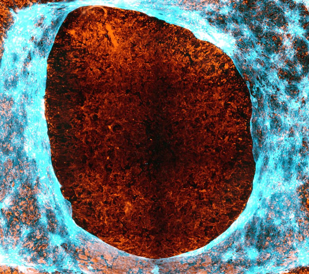
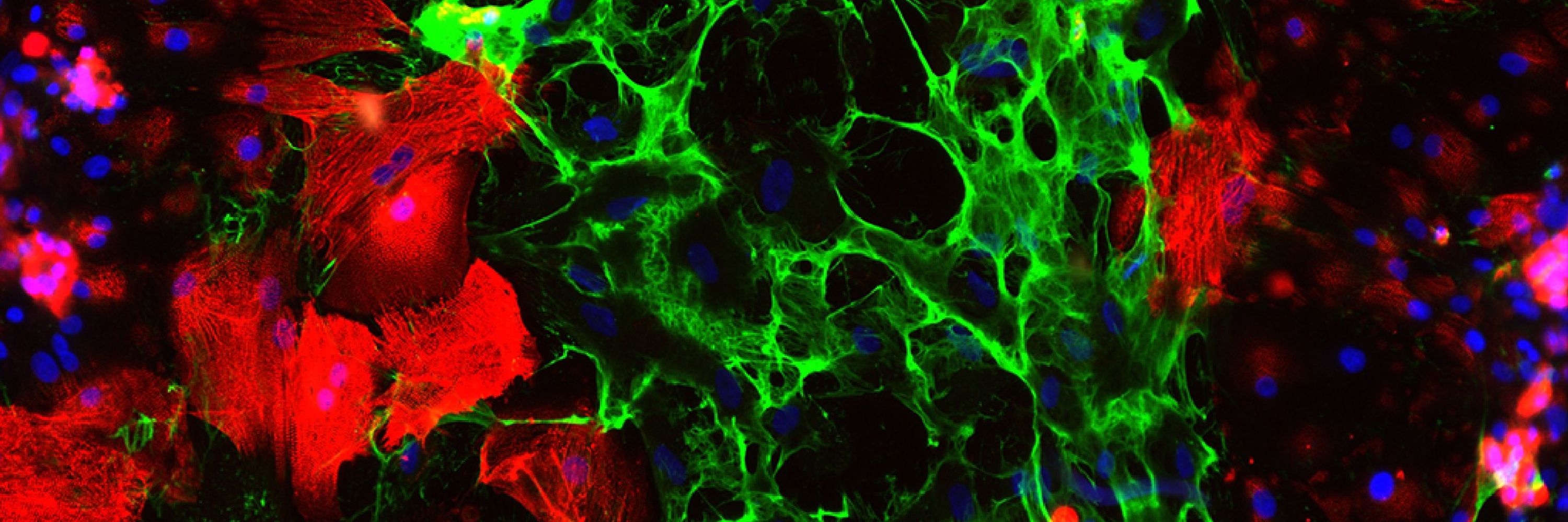
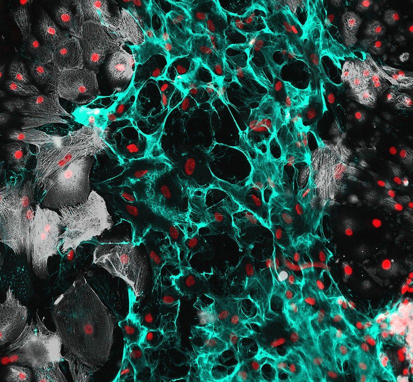
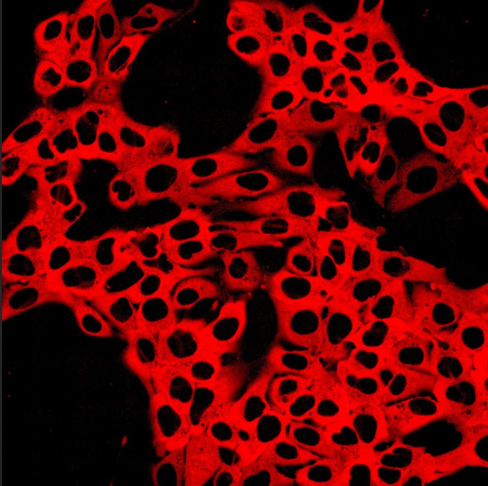
 #FluorescenceFriday
Posts Media Videos Starter Packs
Pinned
benjaminbjohnson.bsky.social
Hello #BlueSky! Figured my first post had to be a #FluorescenceFriday 🧪 I work on #hiPSC cardiac disease modeling ❤️

Image shows the initial collision and fusion of a #Cardioid (right) with a blood vessel organoid (left) as part of a fun collaboration with @mvilacuenca.bsky.social!
Confocal microscopy shows a mini 3D heart (Cardioid) as a sphere full of wavy orange, reds, and cyan in the process of colliding and merging with a blood vessel organoid filled with deep reds and dark blues.
benjaminbjohnson.bsky.social
Sure thing, you’re added!
benjaminbjohnson.bsky.social
Already have you in Cathy 😉
benjaminbjohnson.bsky.social
You’re in my friend 👍🏻
benjaminbjohnson.bsky.social
Sure, you’re in! 👍🏻
benjaminbjohnson.bsky.social
Absolutely, you’re in! 👍🏻
benjaminbjohnson.bsky.social
How to fix a broken #heart 💔->❤️

#Proteoglycan rich #ECMatrix scar tissue is laid by migratory cells after a cardiac scratch wound with #hiPSC #Cardiomyocytes.

Happy #Fluorescencefriday #BlueSky & #CardioSky!

@ablanchasensio.bsky.social 👀
#Science 💥🧪 #Microscopy 🔬
Cardiac scratch wound assay to show the role of ECM in wound healing. HiPSC Cardiomyocytes in white on the boarders with a rich ECM scar tissues (cyan) through the middle with a simple nuclear stain (red) highlighting all cells.
Reposted by Benjamin B Johnson
mag2art.bsky.social
DNA during cell death and cell division.
#Science #Biology #CellBiology #microscopy
benjaminbjohnson.bsky.social
Hi James, would love to be added thanks!
Reposted by Benjamin B Johnson
ablanchasensio.bsky.social
Here is my 2nd #FluorescenceFriday post! 🧪 #hiPSCs expressing jRCaMP1b, a genetically-encoded Ca2+ indicator 👻. This reporter allows us to record intracellular Ca2+ in hiPSC-derived #cardiomyocytes and cardioids (videos in the comments) #microscopy 🔬 #Science #Biology @benjaminbjohnson.bsky.social
Genetically-modified hiPSCs expressing the genetically-encoded calcium indicator jRCaMP1b with a nuclear export signal (NES). This reporter is used to track calcium dynamics in live cells. It combines a red fluorescent protein with calmodulin and a calcium-binding domain, changing fluorescence intensity upon calcium binding. Its red-shifted emission is ideal for imaging in complex tissues or dual-color experiments, making it valuable for studying calcium-mediated processes.
A single copy of this reporter was integrated using the STRAIGHT-IN platform at the AAVS1 locus. See below!
https://doi.org/10.1016/j.crmeth.2022.100300
https://doi.org/10.1101/2024.10.17.616637
Reposted by Benjamin B Johnson
maikbischoff.bsky.social
There are so many #morphogenesis researchers here!
My first starter pack is full!
Here is Part 2: go.bsky.app/K6YYKeC

#devbio #cellbio #science

Tell me if I should add you but try to make sure, that you're not already in Part 1.
I cannot cross-reference everyone 😅

Part 1: go.bsky.app/RzDD9or
benjaminbjohnson.bsky.social
Hi, could I be added to your pack thanks!
benjaminbjohnson.bsky.social
Hey Jeff, would love to be added to your pack thanks!
benjaminbjohnson.bsky.social
Hey, would love to be added! I work on hPSC cardiac systems and disease models ❤️
benjaminbjohnson.bsky.social
Hey, would love to be added thanks! 😀
benjaminbjohnson.bsky.social
Hey, would love to be added to your pack thanks! ❤️
benjaminbjohnson.bsky.social
Hey, can I be added please and thanks! 😀
benjaminbjohnson.bsky.social
Hey! Would love to be added to this pack, thanks! ❤️
benjaminbjohnson.bsky.social
Dedicating this #Fluorescencefriday to the #ECMatrix lovers. Was trialing some ECM staining methods on a scratch wound and love the HABP staining looking almost like flames 🔥🧪🔬

Lets see what you got, @ablanchasensio.bsky.social 😉
Cyan cellular extracellular matrix fibers are highlighted laying over the top of the orange hyaluronan secreted matrix after a scratch would assay.
benjaminbjohnson.bsky.social
Absolutely, you’re in! 👍🏻