@bhadurilab.bsky.social
320 followers 520 following 30 posts
Posts Media Videos Starter Packs
bhadurilab.bsky.social
Special thanks to our amazing collaborators:
Nathanson Lab, Prins Lab, UCLA Neurosurgery, as well as the members of the Bhaduri Lab who have provided advice, edits, and moral support throughout. We look forward to hearing what the community thinks!
bhadurilab.bsky.social
💡iHOTT is a human, patient-specific, matched platform to study tumor–immune interactions. It recapitulates key features of tumor-immune interactions, offering a new tool for immunotherapy discovery.

📄Paper: biorxiv.org/content/10.1...
🌍Data: cells.ucsc.edu?ds=ihott
bhadurilab.bsky.social
🔗Were there shared antigens across patients? We used GLIPH2 to find out. The short answer: not really. Most T cell clonotypes were private to an individual tumor. This tells us something important: GBM’s immune landscape is highly personalized *and* iHOTT robustly captures that.
bhadurilab.bsky.social
To understand the T cell response, we performed TCR sequencing on treated samples. Clonal diversity increased & new clones emerged 🌈. These novel clones were driven by CD4 T-cells and expressed TCF7, a marker for stem-like T cells that has been linked to improved outcomes.
bhadurilab.bsky.social
But MOST interestingly ⭐, when we benchmarked iHOTT against patients treated with pembrolizumab as part of a clinical trial, we noted the *exact same* cell type shifts, validating that iHOTT recapitulates patient biology. This was an exciting finding ‼️
bhadurilab.bsky.social
We treated iHOTT with pembrolizumab 💊to understand the effects of immunotherapy. Interestingly, we noted expansion of CD4+ T cells and B cells and enrichment pathways reflecting immune activation. The cytokine profile also changed with increases in IL15, IL25, and IL17F.
bhadurilab.bsky.social
Our model preserved immune and tumor cell types after co-culture. We also saw immune activation and upregulation of GBM-linked cytokines like IL-6, IL-8, IL-10, and G-CSF, notably seen *only* when tumor cells and PBMCs were cultured together.
bhadurilab.bsky.social
Most GBM models miss one of two things:
🛡️ a functional immune system
🧠 a human brain microenvironment

iHOTT has both.
🧫 We co-culture freshly isolated patient tumor cells + matched PBMCs on human cortical organoids. It’s patient-specific, matched, and immunocompetent.
bhadurilab.bsky.social
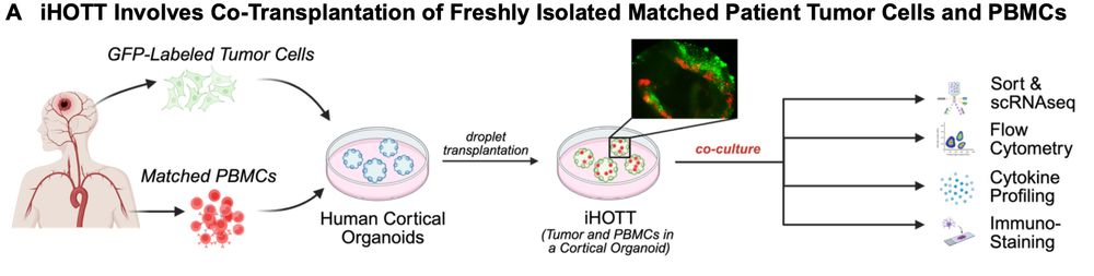
🧵Excited to share our new preprint introducing iHOTT - an autologous tumor-immune co-culture model that captures patient-specific responses in #Glioblastoma

💥Now on
@biorxivpreprint
: biorxiv.org/content/10.1...

Led by Dr. Shivani Baisiwala, Neurosurgery Resident in the lab
biorxiv.org
bhadurilab.bsky.social
A huge thank you to the amazing collaborators and members of the Bhaduri Lab for their contributions to this study! Antoni Martija, @prnano9, @Jalbsoto, Dan Jaklic, @MilJessenya, Rista White, @JacquiMMartn1, Dakshesh Rana, @GeschwindLab
bhadurilab.bsky.social
Taken together, our findings suggest that the early entry of thalamocortical afferents in human and nonhuman primate cortex mediates communication with cortical progenitors. This demonstrates that the human thalamus actively sculpts cortical architecture during early development! 🧩
bhadurilab.bsky.social
Leveraging the tractability of the organoid model, we knocked out thalamic NRXN1 to show that when disrupted, thalamic axons fail to form stable contacts with cortical outer radial glia, leading to impaired upper layer neurogenesis.
bhadurilab.bsky.social
Our key player: NRXN1, a cell adhesion molecule expressed by thalamic neurons, which directly contacts outer radial glia. 🤝 We identify NRXN1-NLGN1 gene expression and protein colocalization along thalamocortical afferents and outer radial glia in the assembloid and human primary cortical tissue.
bhadurilab.bsky.social
Cre-dependent anterograde tracing demonstrates that thalamocortical axons make physical connections with cortical outer radial glia - a primate-enriched progenitor cell type central to cortical expansion. 🧠 But what types of cell-cell interactions mediate this contact?
bhadurilab.bsky.social
Interestingly, severance ✂️ of the thalamic input midway through neurogenesis actually maintains these increased upper layer neurons. But how is early thalamic input changing the future fate of cortical cell types? Our work indicates communication with progenitors may be the answer…
bhadurilab.bsky.social
Utilizing the human thalamocortical assembloid model, we show that thalamic input increases the generation of upper layer cortical neurons compared to cortex-only controls. 🧫 This work shows extrinsic cues like thalamic input can shape intrinsic cortical programs.
bhadurilab.bsky.social
Excited to present our new preprint led by @claudianguyen95 uncovering how thalamic input shapes human cortical development! We discover that thalamic axons promote the generation of upper layer cortical neurons through NRXN1-mediated contacts with outer radial glia. www.biorxiv.org/content/10.1...
bhadurilab.bsky.social
Congratulations @prnano9.bsky.social!!! We are thrilled that this paper is out and hope everyone enjoys using the datasets and meta-atlas tools!!
prnano9.bsky.social
It’s out! The first paper from my postdoc – and first from the @bhadurilab.bsky.social – is now live @natneuro.nature.com . 🧠✨

Using a new meta-atlas generation strategy, we identified functional gene networks that more fully explain how cell types are formed in the human cortex. (1/13)
natneuro.nature.com
Parallel meta-analyses of the human cortex in development (seven datasets) and adulthood (16 datasets) generated over 500 gene co-expression networks that can describe mechanisms of cortical development, centering on peak stages of neurogenesis 🧠🧪

www.nature.com/articles/s41...
bhadurilab.bsky.social
We hope this dataset including metabolomics and scRNA-seq data will be useful to others who are interested in metabolism and cortical cell fates! Shoutout to @madelineandrews.bsky.social and all of our collaborators and the @uclastemcell.bsky.social for contributions and input on this project.
bhadurilab.bsky.social
This led us to a model that indicates that glycolysis, through PPP, seems to restrict radial glia from generating oRGs, astrocytes, and inhibitory neurons too early in development. This may be why inhibition of glycolysis and PPP led to an increase of these cell types.
bhadurilab.bsky.social
To test if PPP was guiding radial glia cell fate, we (painfully) extracted EGFP labeled radial from organoids and knocked down PPP genes. Indeed, same story. More oRGs, astrocytes, and inhibitory neurons paired with a decrease in PPP metabolite abundance.
bhadurilab.bsky.social
This was fascinating because PPP also peaks in primary tissue at the same time as glycolysis. Was glycolysis working through the PPP? We tested this by inhibiting PPP with pharmacological inhibition, and also saw an increase in oRGs, astrocytes, and inhibitory neurons.
bhadurilab.bsky.social
Organoids love sugar! Using staining and scRNA-seq, we saw an increase in outer radial glia (oRGs), astrocytes, and inhibitory neurons. When we examined metabolite abundance in this experiment many pathways, including the pentose phosphate pathway (PPP) were downregulated.
bhadurilab.bsky.social
This was intriguing because glycolysis has been a flash point and somewhat controversial in organoids. Is it up in organoids? And if so, does it impact cell fate? We tested this by lowering glucose levels in organoid culture and measuring how cell types changed.
bhadurilab.bsky.social
We saw known changes in metabolites, and discovered many novel dynamics. When comparing organoids to primary tissue, we saw that many metabolites were preserved (80%) but some had opposite trends over development. Some of these were in downstream parts of glycolysis…