


Benedikt Wimmer
@bwmr.net
Using cryo-ET and light microscopy to study bacteria and how they help and hurt us.
PostDoc at the Jacobs-Wagner lab in Stanford. Previously at the Medalia Lab (Zurich) and Chlanda lab (Heidelberg). #teamtomo
PostDoc at the Jacobs-Wagner lab in Stanford. Previously at the Medalia Lab (Zurich) and Chlanda lab (Heidelberg). #teamtomo
- We modelled the active sites of two substrate-specific amylosome enzymes and compared it to the bifunctional Amy16, to derive a structural hypothesis for the observed promiscuity.

December 1, 2025 at 7:45 PM
- We modelled the active sites of two substrate-specific amylosome enzymes and compared it to the bifunctional Amy16, to derive a structural hypothesis for the observed promiscuity.
- In collaboration with the Zeeman lab @ethz.ch we further characterized the products released by Amy16, a key enzyme in RS degradation. This revealed that Amy16 is a true bifunctional amylase and pullulanase, hydrolyzing both α(1,4)- and α(1,6)-linkages - supporting its key role in RS degradation.

December 1, 2025 at 7:45 PM
- In collaboration with the Zeeman lab @ethz.ch we further characterized the products released by Amy16, a key enzyme in RS degradation. This revealed that Amy16 is a true bifunctional amylase and pullulanase, hydrolyzing both α(1,4)- and α(1,6)-linkages - supporting its key role in RS degradation.
- We modelled all amylosome proteins detected in our proteomics sample using AlphaFold3 and built a model of amylosome distribution around the cell wall, matching nicely the densities we observed using cryo-ET.

December 1, 2025 at 7:45 PM
- We modelled all amylosome proteins detected in our proteomics sample using AlphaFold3 and built a model of amylosome distribution around the cell wall, matching nicely the densities we observed using cryo-ET.
(4/5)
By combining in-situ #cryoet and #proteomics, we could assemble an integrative model, which illustrates the remarkable architecture of the amylosome - the complex that allows R. bromii to act as keystone degrader in the gut microbiome.
By combining in-situ #cryoet and #proteomics, we could assemble an integrative model, which illustrates the remarkable architecture of the amylosome - the complex that allows R. bromii to act as keystone degrader in the gut microbiome.
April 3, 2025 at 12:41 PM
(4/5)
By combining in-situ #cryoet and #proteomics, we could assemble an integrative model, which illustrates the remarkable architecture of the amylosome - the complex that allows R. bromii to act as keystone degrader in the gut microbiome.
By combining in-situ #cryoet and #proteomics, we could assemble an integrative model, which illustrates the remarkable architecture of the amylosome - the complex that allows R. bromii to act as keystone degrader in the gut microbiome.
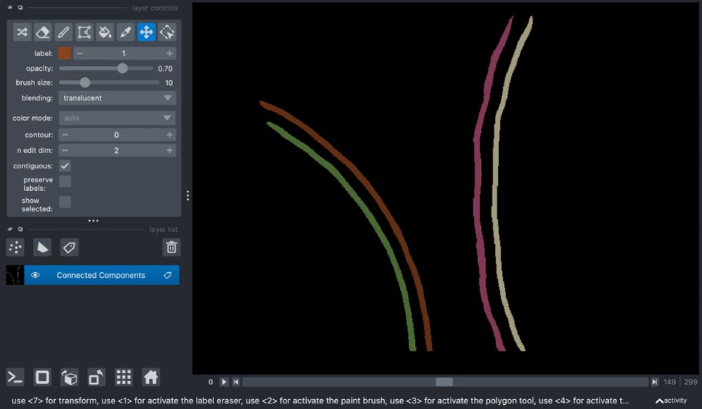
... which can then be written out as binary masks in the mrc format and visualized or used however you like:
February 6, 2025 at 11:02 AM
... which can then be written out as binary masks in the mrc format and visualized or used however you like:
... and allows you to easily create labels containing only the IDs of interest.
February 6, 2025 at 11:02 AM
... and allows you to easily create labels containing only the IDs of interest.
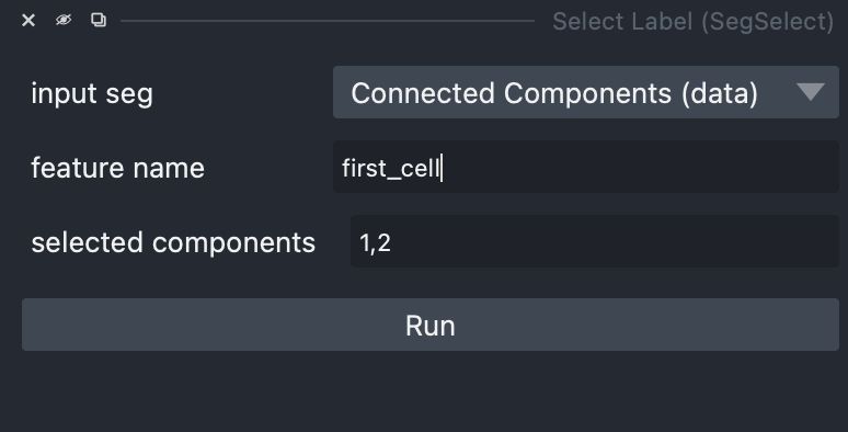
"napari-segselect" automatically opens the "*_segmented.mrc" files from membrain-seg as a label field...
February 6, 2025 at 11:02 AM
"napari-segselect" automatically opens the "*_segmented.mrc" files from membrain-seg as a label field...
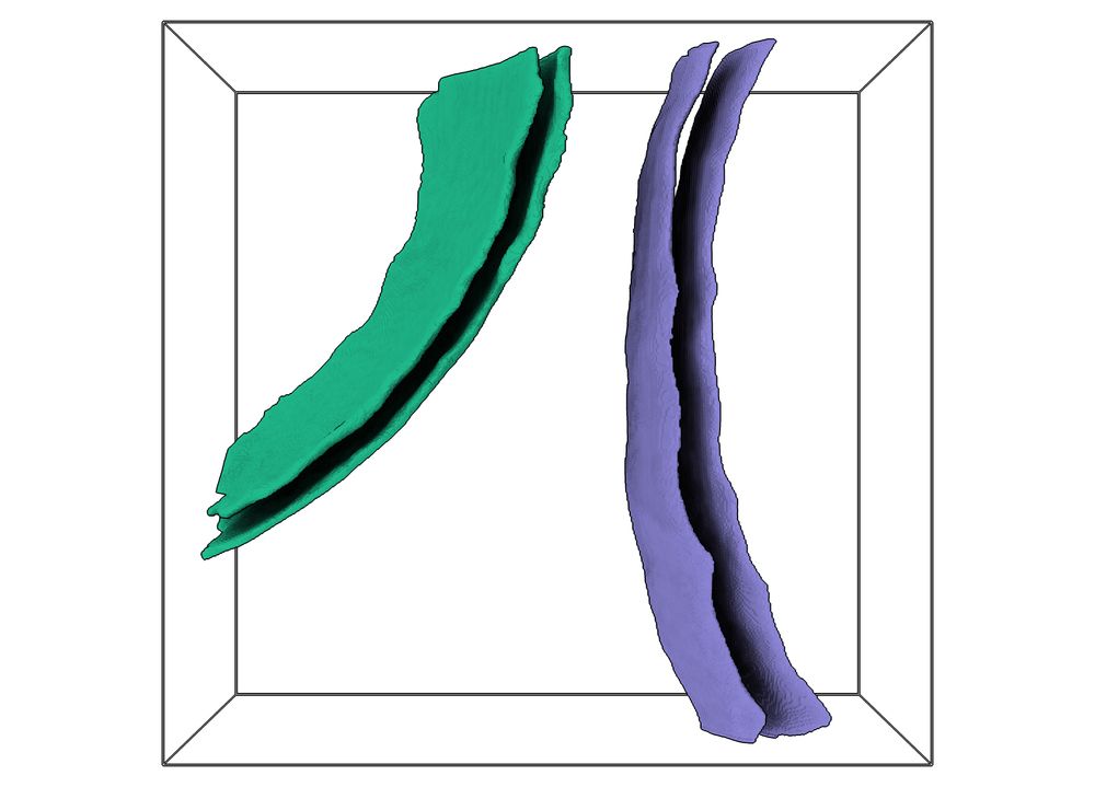
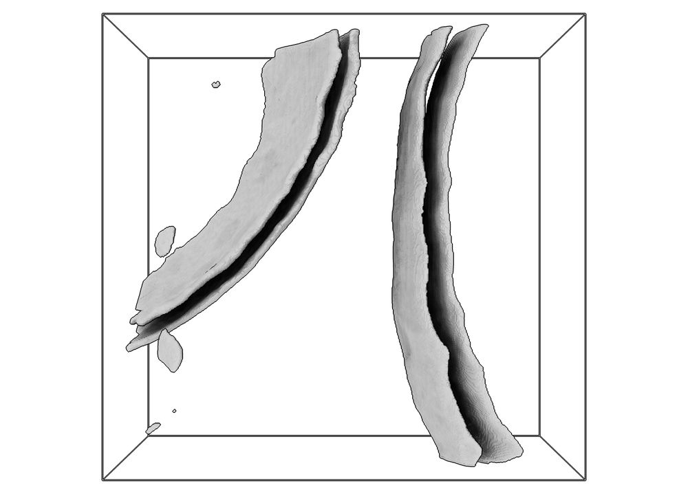
Hey #teamtomo, if you often find yourself using the excellent membrain-seg from @lorenzlamm.bsky.social et al., you might find my napari plugin "napari-segselect" useful. Let's say your tomogram contains the edges of two bacterial cells, each with a membrane and cell wall:
February 6, 2025 at 11:02 AM
Hey #teamtomo, if you often find yourself using the excellent membrain-seg from @lorenzlamm.bsky.social et al., you might find my napari plugin "napari-segselect" useful. Let's say your tomogram contains the edges of two bacterial cells, each with a membrane and cell wall: