Christiana Garros
@chrissygarros.bsky.social
300 followers
120 following
40 posts
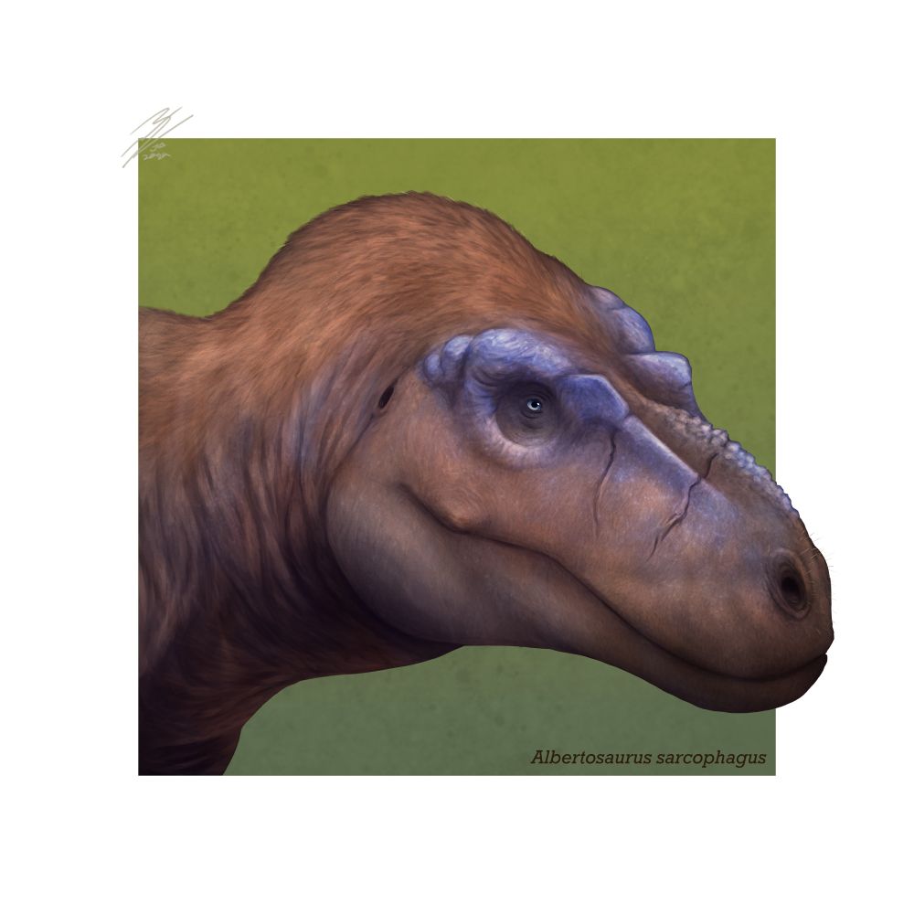
🇨🇦🇧🇷| Edmonton | U of Alberta | MSc student in paleontology studying theropod foot pathologies 🦖 | she/her
Posts
Media
Videos
Starter Packs
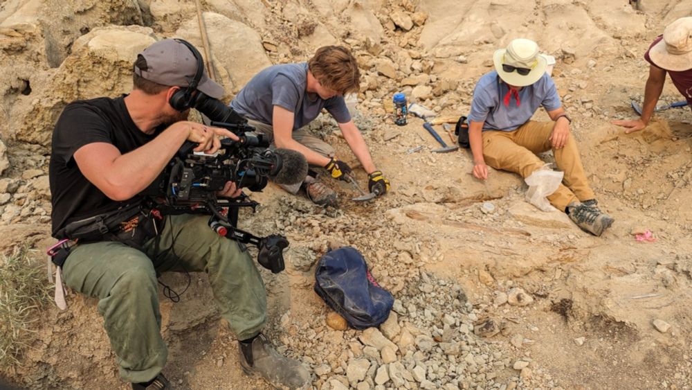
Reposted by Christiana Garros