Eric Surette
@ericsurette.bsky.social
31 followers 45 following 1 posts
Developmental biologist studying the formation and evolution of skeletal shapes 🐟 🦴. He/him/his
Posts Media Videos Starter Packs
Reposted by Eric Surette
aurhin.bsky.social
Are digits modified fins, or evolutionary innovations? Read how we tackled this old question from a new angle🧪
A story with @chasebolt.bsky.social, @homeobox.bsky.social and myself, coordinated by @denisduboule.bsky.social from @college-de-france.fr and published in @nature.com today!
#InHoxWeTrust
hoxd13a reporter in a zebrafish larva
Reposted by Eric Surette
plosbiology.org
What guides fibroblast precursors' migration from the #sclerotome during vertebrate development? @penghuang031.bsky.social &co show that this is driven by #PDGF -mediated chemoattraction, enabling their differentiation into specialized #fibroblast subtypes @plosbiology.org 🧪 plos.io/3HTZ92Y
Top: Transgenic expression of PdgfraΔK-EGFP (cyan) in nkx3.1:Gal4; UAS:NTR-mCherry (magenta) background at 56 hpf. mCherry+ EGFP+ cells are mostly restricted in the dorsal or ventral regions of the trunk. A transverse view of a 50 μm region centered on the dashed line is shown on the right, with the neural tube (nt) and notochord (n) outlined by dotted lines. Bottom: Model for how the migration of sclerotome-derived cells is regulated by chemoattraction mediated by Pdgfab/Pdgfra signaling. (A) In wild-type embryos, nkx3.1+ (cyan) sclerotome-derived cells express the receptor Pdgfra (magenta), while the medial somites express the ligand Pdgfab (orange) in response to Hh signaling. pdgfra+ sclerotome-derived cells undergo extensive migration toward both the notochord in the medial trunk and the fin fold. (B) In pdgfra−/− mutants (pdgframRFP/mRFP and pdgfraref/ref), nkx3.1+ sclerotome-derived cells fail to migrate in any direction and remain in their original location in the ventral and dorsal parts of the somites. (C) In pdgfab−/− embryos, pdgfra+ sclerotome-derived cells fail to migrate toward the notochord, although migration toward the fin fold remains unaffected. (D) In pdgfab−/− embryos with mosaic pdgfab expression, nkx3.1+ sclerotome cells can migrate toward the ectopic pdgfab-expressing cells, either medially around the notochord or ventrally toward the yolk extension. When the pdgfab source is located near or in the notochord, it can completely rescue the migration of nkx3.1+ sclerotome cells toward the notochord. These diagrams depict a 2-somite region located above the yolk extension of a zebrafish embryo at 48 hpf. nt: neural tube; n: notochord.
Reposted by Eric Surette
homeobox.bsky.social
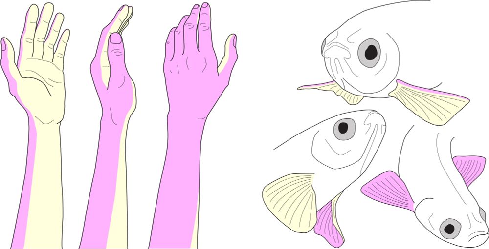
We take for granted that our hands have two sides and can articulate in an endless number of ways. But what about a fish’s fin? Can a fish know something “like the back of its fin,” or have its future told with a fin palm reading? Check out this bluetorial to find out 👇🧪🧬🐟 doi.org/10.1101/2025...
Schematics depicting the dorsal and ventral sides of fins and limbs
ericsurette.bsky.social
Hello Bluesky: Long time listener, first time poster! Excited to share the first publication of my Ph.D. work studying the development of appendage shapes in @plosbiology.org! The zebrafish caudal fin is quite distinctive, and we examine the timing and mechanisms of how its shape might come to be!
plosbiology.org
Caudal fins show great variation in shape between diverse fish species. New research reveals that #developmental cues in embryonic tissues involving the Sonic hedgehog pathway imprint fin #morphology, highlighting a critical window for modulating appendage morphology. 🧪
plos.io/4mydLnA
Brightfield image of an adult zebrafish caudal fin displaying a “truncate” shape and aberrant pigment pattern. This individual was exposed to transient overexpression of transgenic sonic hedgehog a at 2 days post fertilization, resulting in increased growth of rays at the center of the fin and disruption of the normally horizontal pigment stripes. Credit: Eric Surette and Stephanie Robinson.