PLOS Biology
@plosbiology.org
13K followers 7K following 1.3K posts
A leading life science journal that champions high-impact research across all disciplines—from molecules to ecosystems. We offer innovative formats and collaborative editorial support to ensure your work achieves its full scientific impact.
Posts Media Videos Starter Packs
Pinned
plosbiology.org
PLOS has issued a statement on recent US Executive Orders and scientific integrity.

We are determined to stand firmly behind our mission, our values and our principles, and against any attempt at censorship or undermining of the core principles of scientific inquiry.

plos.io/3D4O8cH
PLOS statement on recent US Executive Orders and scientific integrity - The Official PLOS Blog
Since its founding over twenty five years ago PLOS has been dedicated to advancing open science, ensuring that knowledge is accessible to…
plos.io
plosbiology.org
Sleep is often only investigated from a single dimension. @bttyeo.bsky.social &co identify 5 sleep-biopsychosocial profiles that link self-reported #sleep patterns to variability in #health, #cognition & #lifestyle factors in 770 healthy young adults @plosbiology.org 🧪 plos.io/42tRXSc
Canonical correlation analysis reveals five sleep-biopsychosocial profiles (LCs). Scatter plots showing correlations between biopsychosocial and sleep canonical scores. Each dot represents a different participant. The inset shows the null distribution of canonical correlations obtained by permutation testing; note that the null distribution is not centered at zero. The dashed line indicates the actual canonical correlation computed for each LC. The distribution of sleep (top) and biopsychosocial (right) canonical scores is shown on rain cloud plots.
plosbiology.org
How does attention shape #AuditoryProcessing? This study shows that #attention enhances sound encoding only at the cortical level, not in #auditory periphery or #brainstem, so humans’ ability to focus on a single voice in noise relies on cortical mechanisms @plosbiology.org 🧪 plos.io/4pY5L1f
Subcortical sound encoding is not affected by selective attention, while cortical sound encoding is. Top left: The CAP to the attended (red) and unattended (black) speech for the 12 subjects with the eardrum electrode. Bottom left: The ABR for all 28 subjects when attended (red) or unattended (black). No significant differences were observed between conditions for either the CAP or ABR. Shaded regions represent SEM. Right: Cortical responses to attended (top) and unattended (middle) stimuli. Bottom right: The difference waveform and scalp topographies at the selected time points. Asterisks indicate the electrodes that were significantly different across conditions at that time.
plosbiology.org
During #development, distal visceral #endoderm (DVE) cells migrate to shape A-P axis specification. @m-stower.bsky.social @srinivas-lab.bsky.social &co reveal the role of Ca2+ dynamics in this, showing that inhibition of #calcium transients causes DVE arrest @plosbiology.org 🧪 plos.io/4nHWxF4
Epiblast Ca2+ waves propagate from the apical to basal domain. The images show selected time points from movies of E5.5 GCaMP6f embryos imaged using a ZEISS lattice light-sheet 7 microscope. Top row: An example of an epiblast wave involving a large proportion of the tissue captured by imaging a volume of the embryo at 20-s intervals. A total of 9/16 embryos showed at least one epiblast Ca2+ wave. Bottom row: Faster imaging at 5-s interval revealed that the Ca2+ wave in the epiblast can be preceded by a transient in a neighboring VE cell—this was observed in 5/19 events from 9 embryos (white arrowheads).
plosbiology.org
How does attention shape #AuditoryProcessing? This study shows that #attention enhances sound encoding only at the cortical level, not in #auditory periphery or #brainstem, so humans’ ability to focus on a single voice in noise relies on cortical mechanisms @plosbiology.org 🧪 plos.io/4pY5L1f
Subcortical sound encoding is not affected by selective attention, while cortical sound encoding is. Top left: The CAP to the attended (red) and unattended (black) speech for the 12 subjects with the eardrum electrode. Bottom left: The ABR for all 28 subjects when attended (red) or unattended (black). No significant differences were observed between conditions for either the CAP or ABR. Shaded regions represent SEM. Right: Cortical responses to attended (top) and unattended (middle) stimuli. Bottom right: The difference waveform and scalp topographies at the selected time points. Asterisks indicate the electrodes that were significantly different across conditions at that time.
Reposted by PLOS Biology
mitisbio.bsky.social
Our calcium paper is out in PLOS Biology!
Check it out! #Calcium #Embryo #Development
plosbiology.org
During #development, distal visceral #endoderm (DVE) cells migrate to shape A-P axis specification. @m-stower.bsky.social @srinivas-lab.bsky.social &co reveal the role of Ca2+ dynamics in this, showing that inhibition of #calcium transients causes DVE arrest @plosbiology.org 🧪 plos.io/4nHWxF4
Epiblast Ca2+ waves propagate from the apical to basal domain. The images show selected time points from movies of E5.5 GCaMP6f embryos imaged using a ZEISS lattice light-sheet 7 microscope. Top row: An example of an epiblast wave involving a large proportion of the tissue captured by imaging a volume of the embryo at 20-s intervals. A total of 9/16 embryos showed at least one epiblast Ca2+ wave. Bottom row: Faster imaging at 5-s interval revealed that the Ca2+ wave in the epiblast can be preceded by a transient in a neighboring VE cell—this was observed in 5/19 events from 9 embryos (white arrowheads).
plosbiology.org
How does attention shape #AuditoryProcessing? This study shows that #attention enhances sound encoding only at the cortical level, not in #auditory periphery or #brainstem, so humans’ ability to focus on a single voice in noise relies on cortical mechanisms @plosbiology.org 🧪 plos.io/4pY5L1f
Subcortical sound encoding is not affected by selective attention, while cortical sound encoding is. Top left: The CAP to the attended (red) and unattended (black) speech for the 12 subjects with the eardrum electrode. Bottom left: The ABR for all 28 subjects when attended (red) or unattended (black). No significant differences were observed between conditions for either the CAP or ABR. Shaded regions represent SEM. Right: Cortical responses to attended (top) and unattended (middle) stimuli. Bottom right: The difference waveform and scalp topographies at the selected time points. Asterisks indicate the electrodes that were significantly different across conditions at that time.
plosbiology.org
During #development, distal visceral #endoderm (DVE) cells migrate to shape A-P axis specification. @m-stower.bsky.social @srinivas-lab.bsky.social &co reveal the role of Ca2+ dynamics in this, showing that inhibition of #calcium transients causes DVE arrest @plosbiology.org 🧪 plos.io/4nHWxF4
Epiblast Ca2+ waves propagate from the apical to basal domain. The images show selected time points from movies of E5.5 GCaMP6f embryos imaged using a ZEISS lattice light-sheet 7 microscope. Top row: An example of an epiblast wave involving a large proportion of the tissue captured by imaging a volume of the embryo at 20-s intervals. A total of 9/16 embryos showed at least one epiblast Ca2+ wave. Bottom row: Faster imaging at 5-s interval revealed that the Ca2+ wave in the epiblast can be preceded by a transient in a neighboring VE cell—this was observed in 5/19 events from 9 embryos (white arrowheads).
Reposted by PLOS Biology
kiraposkanzer.bsky.social
Thanks to @ashleyingiosi.bsky.social for this thoughtful primer about our recent astrocyte/sleep paper!
plosbiology.org
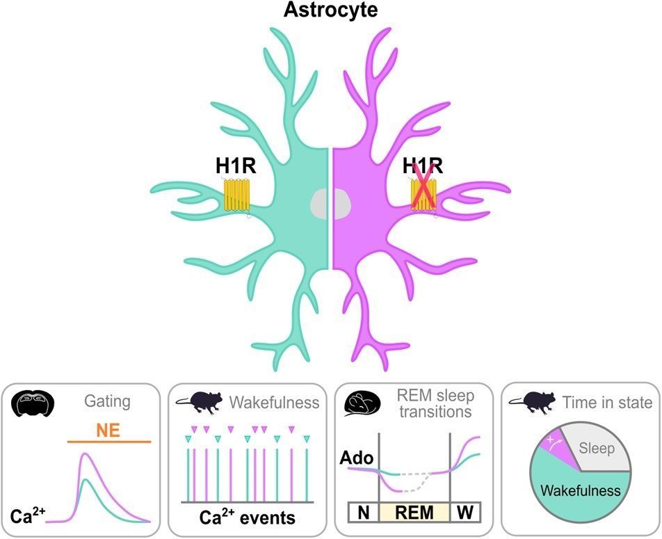
Astrocytes help control #sleep & arousal. @ashleyingiosi.bsky.social explores a @plosbiology.org study showing that wake-promoting #histamine affects how #astrocytes respond to other signals, reducing awake time via astroglial histamine-1-receptors 🧪 Paper: plos.io/4gRj9Ap Primer: plos.io/47at1Sj
Astroglial H1Rs influence neuromodulatory integration and arousal.

How histamine modulates arousal via astrocytes was investigated in brain slices and mice with (left; green) and without (right; purple) histamine-1-receptor (H1R) expression in astrocytes of the primary visual cortex (V1). Panels below depict, from left to right, that H1R expression in V1 astrocytes: (1) attenuates astroglial calcium (Ca²⁺) responses to norepinephrine (NE) in brain slices; (2) suppresses astroglial Ca²⁺ events during wakefulness; (3) restricts the dynamic range of extracellular adenosine (Ado) around rapid eye movement (REM) sleep transitions (N, non-rapid eye movement sleep; W, wakefulness); and (4) reduces time spent in wakefulness.
plosbiology.org
Nicotine exposure elicits diverse behavioral changes. This study shows that in #Celegans, chronic #nicotine exposure couples reversal behaviors to rhythmic body contractions by modulating the sensitivity of a mechanosensory-motor circuit @plosbiology.org 🧪 plos.io/4o2CWyS
Top: Nicotine up-regulates ACR-16 expression in AVA. Using fluorescently-tagged ACR-16 based on split GFP reconstitution, the authors inserted three copies of the spGFP11 (split GFP11) sequence into the acr-16::aid::Scarlet locus via CRISPR/Cas9 (ACR-16-AID-Scarlet-spGFP11) and expressed the complementary spGFP1-10 (split GFP1-10) moiety in the AVA to generate AVA specific ACR-16::GFP transgenic line. Scale bar, 10 µm. Bottom: Working model. Nicotine significantly induces abnormal DMP-reversal coupling response in Caenorhabditis elegans. An unexpected mechanosensory neural pathway involving TRN-AVA circuit mediates this response. Key mechanisms include the nicotine-induced upregulation of ACR-16 nAChRs in AVA and nicotine-strengthened signal transmission within TRN-AVA circuit. TRNs may sense internal body contractions associated with the DMP, abnormally activating sensitized command interneurons to drive reversals. This finding underscores an unexpected functional integration induced by nicotine exposure.
plosbiology.org
Astrocytes help control #sleep & arousal. @ashleyingiosi.bsky.social explores a @plosbiology.org study showing that wake-promoting #histamine affects how #astrocytes respond to other signals, reducing awake time via astroglial histamine-1-receptors 🧪 Paper: plos.io/4gRj9Ap Primer: plos.io/47at1Sj
Astroglial H1Rs influence neuromodulatory integration and arousal.

How histamine modulates arousal via astrocytes was investigated in brain slices and mice with (left; green) and without (right; purple) histamine-1-receptor (H1R) expression in astrocytes of the primary visual cortex (V1). Panels below depict, from left to right, that H1R expression in V1 astrocytes: (1) attenuates astroglial calcium (Ca²⁺) responses to norepinephrine (NE) in brain slices; (2) suppresses astroglial Ca²⁺ events during wakefulness; (3) restricts the dynamic range of extracellular adenosine (Ado) around rapid eye movement (REM) sleep transitions (N, non-rapid eye movement sleep; W, wakefulness); and (4) reduces time spent in wakefulness.
plosbiology.org
Evolution of fungal mating systems: Study of #Cryptococcus & Kwoniella by @marcoacoelho.bsky.social @marciadavidpalma.bsky.social &co shows that MAT locus fusions & recombination suppression drive diversity of #SexualReproduction in #fungi @plosbiology.org 🧪 plos.io/479579M
Top: Micrographs showing hyphal filaments extending from colony peripheries in a sexual cross between CBS8507 and CBS10435 after 2 weeks of incubation on CMA and V8 pH 5 media, in the dark, at room temperature. Insets depict two types of basidia: globose (a–c) and lageniform (b–d). Black arrows indicate clusters of cells emerging near basidia (either at the surface or embedded), scored as potential meiotic progeny (basidiospores). Bottom: Schematic representation of key chromosomal rearrangements and evolutionary events underlying transitions from tetrapolar to bipolar and pseudobipolar mating configurations. The phylogenetic tree highlights inferred changes in MAT locus structure, with color-coded branches representing both extant and reconstructed MAT configurations. Dashed lines indicate unresolved phylogenetic relationships among clades A, B, and C. Insets summarize distinct evolutionary stages, including: the ancestral organization with unlinked and compact P/R and HD loci (1); small-scale expansion of the P/R locus in Kwoniella (2); two independent large-scale expansions of the P/R locus in Cryptococcus (5); relocation of P/R and HD loci onto the same chromosome in Kwoniella resulting in either pseudobipolar (3) or fused (4) MAT configurations; and independent HD-P/R fusion events with HD gene loss in Cryptococcus pathogens and Cryptococcus sp. 3 (6). These transitions illustrate the diverse pathways by which chromosomal rearrangements (CRs) have shaped MAT locus architecture in these fungal lineages.
Reposted by PLOS Biology
droutledge.bsky.social
In our latest Editorial, PLOS Medicine Executive Editor Helen Lumbard and I call for the scientific community to tackle research fraud and paper mills head on by creating robust systems of data sharing and transparency 🧪 #PLOSMedicine #datasharing #openscience
@plos.org

📄➡️ plos.io/4nDKQ2c
Open science and transparency are our strongest tools in the fight against fraudulent publishing activities
PLOS Medicine Executive Editor, Helen Lumbard, and Senior Editor, Front Section, Daniel Routledge, call for the scientific community to tackle research fraud and paper mills head on by creating robust...
journals.plos.org
plosbiology.org
Evolution of fungal mating systems: Study of #Cryptococcus & Kwoniella by @marcoacoelho.bsky.social @marciadavidpalma.bsky.social &co shows that MAT locus fusions & recombination suppression drive diversity of #SexualReproduction in #fungi @plosbiology.org 🧪 plos.io/479579M
Top: Micrographs showing hyphal filaments extending from colony peripheries in a sexual cross between CBS8507 and CBS10435 after 2 weeks of incubation on CMA and V8 pH 5 media, in the dark, at room temperature. Insets depict two types of basidia: globose (a–c) and lageniform (b–d). Black arrows indicate clusters of cells emerging near basidia (either at the surface or embedded), scored as potential meiotic progeny (basidiospores). Bottom: Schematic representation of key chromosomal rearrangements and evolutionary events underlying transitions from tetrapolar to bipolar and pseudobipolar mating configurations. The phylogenetic tree highlights inferred changes in MAT locus structure, with color-coded branches representing both extant and reconstructed MAT configurations. Dashed lines indicate unresolved phylogenetic relationships among clades A, B, and C. Insets summarize distinct evolutionary stages, including: the ancestral organization with unlinked and compact P/R and HD loci (1); small-scale expansion of the P/R locus in Kwoniella (2); two independent large-scale expansions of the P/R locus in Cryptococcus (5); relocation of P/R and HD loci onto the same chromosome in Kwoniella resulting in either pseudobipolar (3) or fused (4) MAT configurations; and independent HD-P/R fusion events with HD gene loss in Cryptococcus pathogens and Cryptococcus sp. 3 (6). These transitions illustrate the diverse pathways by which chromosomal rearrangements (CRs) have shaped MAT locus architecture in these fungal lineages.
plosbiology.org
Astrocytes help control #sleep & arousal. @ashleyingiosi.bsky.social explores a @plosbiology.org study showing that wake-promoting #histamine affects how #astrocytes respond to other signals, reducing awake time via astroglial histamine-1-receptors 🧪 Paper: plos.io/4gRj9Ap Primer: plos.io/47at1Sj
Astroglial H1Rs influence neuromodulatory integration and arousal.

How histamine modulates arousal via astrocytes was investigated in brain slices and mice with (left; green) and without (right; purple) histamine-1-receptor (H1R) expression in astrocytes of the primary visual cortex (V1). Panels below depict, from left to right, that H1R expression in V1 astrocytes: (1) attenuates astroglial calcium (Ca²⁺) responses to norepinephrine (NE) in brain slices; (2) suppresses astroglial Ca²⁺ events during wakefulness; (3) restricts the dynamic range of extracellular adenosine (Ado) around rapid eye movement (REM) sleep transitions (N, non-rapid eye movement sleep; W, wakefulness); and (4) reduces time spent in wakefulness.
plosbiology.org
Nicotine exposure elicits diverse behavioral changes. This study shows that in #Celegans, chronic #nicotine exposure couples reversal behaviors to rhythmic body contractions by modulating the sensitivity of a mechanosensory-motor circuit @plosbiology.org 🧪 plos.io/4o2CWyS
Top: Nicotine up-regulates ACR-16 expression in AVA. Using fluorescently-tagged ACR-16 based on split GFP reconstitution, the authors inserted three copies of the spGFP11 (split GFP11) sequence into the acr-16::aid::Scarlet locus via CRISPR/Cas9 (ACR-16-AID-Scarlet-spGFP11) and expressed the complementary spGFP1-10 (split GFP1-10) moiety in the AVA to generate AVA specific ACR-16::GFP transgenic line. Scale bar, 10 µm. Bottom: Working model. Nicotine significantly induces abnormal DMP-reversal coupling response in Caenorhabditis elegans. An unexpected mechanosensory neural pathway involving TRN-AVA circuit mediates this response. Key mechanisms include the nicotine-induced upregulation of ACR-16 nAChRs in AVA and nicotine-strengthened signal transmission within TRN-AVA circuit. TRNs may sense internal body contractions associated with the DMP, abnormally activating sensitized command interneurons to drive reversals. This finding underscores an unexpected functional integration induced by nicotine exposure.
plosbiology.org
Astrocytes help control #sleep & arousal. @ashleyingiosi.bsky.social explores a @plosbiology.org study showing that wake-promoting #histamine affects how #astrocytes respond to other signals, reducing awake time via astroglial histamine-1-receptors 🧪 Paper: plos.io/4gRj9Ap Primer: plos.io/47at1Sj
Astroglial H1Rs influence neuromodulatory integration and arousal.

How histamine modulates arousal via astrocytes was investigated in brain slices and mice with (left; green) and without (right; purple) histamine-1-receptor (H1R) expression in astrocytes of the primary visual cortex (V1). Panels below depict, from left to right, that H1R expression in V1 astrocytes: (1) attenuates astroglial calcium (Ca²⁺) responses to norepinephrine (NE) in brain slices; (2) suppresses astroglial Ca²⁺ events during wakefulness; (3) restricts the dynamic range of extracellular adenosine (Ado) around rapid eye movement (REM) sleep transitions (N, non-rapid eye movement sleep; W, wakefulness); and (4) reduces time spent in wakefulness.
plosbiology.org
Evolution of fungal mating systems: Study of #Cryptococcus & Kwoniella by @marcoacoelho.bsky.social @marciadavidpalma.bsky.social &co shows that MAT locus fusions & recombination suppression drive diversity of #SexualReproduction in #fungi @plosbiology.org 🧪 plos.io/479579M
Top: Micrographs showing hyphal filaments extending from colony peripheries in a sexual cross between CBS8507 and CBS10435 after 2 weeks of incubation on CMA and V8 pH 5 media, in the dark, at room temperature. Insets depict two types of basidia: globose (a–c) and lageniform (b–d). Black arrows indicate clusters of cells emerging near basidia (either at the surface or embedded), scored as potential meiotic progeny (basidiospores). Bottom: Schematic representation of key chromosomal rearrangements and evolutionary events underlying transitions from tetrapolar to bipolar and pseudobipolar mating configurations. The phylogenetic tree highlights inferred changes in MAT locus structure, with color-coded branches representing both extant and reconstructed MAT configurations. Dashed lines indicate unresolved phylogenetic relationships among clades A, B, and C. Insets summarize distinct evolutionary stages, including: the ancestral organization with unlinked and compact P/R and HD loci (1); small-scale expansion of the P/R locus in Kwoniella (2); two independent large-scale expansions of the P/R locus in Cryptococcus (5); relocation of P/R and HD loci onto the same chromosome in Kwoniella resulting in either pseudobipolar (3) or fused (4) MAT configurations; and independent HD-P/R fusion events with HD gene loss in Cryptococcus pathogens and Cryptococcus sp. 3 (6). These transitions illustrate the diverse pathways by which chromosomal rearrangements (CRs) have shaped MAT locus architecture in these fungal lineages.
plosbiology.org
Nicotine exposure elicits diverse behavioral changes. This study shows that in #Celegans, chronic #nicotine exposure couples reversal behaviors to rhythmic body contractions by modulating the sensitivity of a mechanosensory-motor circuit @plosbiology.org 🧪 plos.io/4o2CWyS
Top: Nicotine up-regulates ACR-16 expression in AVA. Using fluorescently-tagged ACR-16 based on split GFP reconstitution, the authors inserted three copies of the spGFP11 (split GFP11) sequence into the acr-16::aid::Scarlet locus via CRISPR/Cas9 (ACR-16-AID-Scarlet-spGFP11) and expressed the complementary spGFP1-10 (split GFP1-10) moiety in the AVA to generate AVA specific ACR-16::GFP transgenic line. Scale bar, 10 µm. Bottom: Working model. Nicotine significantly induces abnormal DMP-reversal coupling response in Caenorhabditis elegans. An unexpected mechanosensory neural pathway involving TRN-AVA circuit mediates this response. Key mechanisms include the nicotine-induced upregulation of ACR-16 nAChRs in AVA and nicotine-strengthened signal transmission within TRN-AVA circuit. TRNs may sense internal body contractions associated with the DMP, abnormally activating sensitized command interneurons to drive reversals. This finding underscores an unexpected functional integration induced by nicotine exposure.
Reposted by PLOS Biology
zebrafishrock.bsky.social
#ZebrafishFunFacts: As the most-cited #zebrafish cancer research paper, Amsterdam et al screened hundreds of heterozygous lines for embryonic lethal mutations & found elevated cancer incidence in ribosomal genes. This previously underappreciated finding was published @plosbiology.org 21 years ago. 🧪
Figure 1. Spindle Cell Tumors Resembling MPNSTs in Zebrafish Heterozygous for Mutations in RP Genes
(A and B) Fish with apparent masses, as indicated by the arrows, or other evident pathology were selected for histological analysis: (A) a hi2582 fish, (B) a hi1034B fish. (C–H) Histopathology of representative tumors stained with hematoxylin and eosin reveals patterns consistent with the diagnosis of MPNST in hi10 fish (C and D), hi1974 fish (E–G), and hi1807 fish (H). (D) A large tumor with central necrosis is seen emanating from the optic nerve (n) (e, eye) (20×).
plosbiology.org
What's the function of #histamine -1-receptors (H1Rs) on cortical astrocytes? @kiraposkanzer.bsky.social & co show that astrocytic H1R signaling modulates Ca2+ activity, extracellular adenosine & REM #sleep, revealing a key #astrocyte role in sleep-wake regulation @plosbiology.org 🧪 plos.io/4gRj9Ap
Top: Example confocal micrograph showing astrocyte expression of S100β (green), Cre-RFP (magenta), and H1R mRNA (cyan). Left: summed z-projection of all three confocal channels. Middle: RNAscope detected H1R mRNA with white and yellow arrows indicating example WT (S100β+/RFP−/H1R+) and cKO (RFP+/H1R−) soma regions of interest (ROIs), respectively. Right: Cre-RFP expression (top, magenta) and S100β expression (bottom, green). Scale bars = 10 μm. Bottom: Example confocal micrograph showing astrocytic Cre-RFP (magenta), neuronal NeuN (green), and nuclear marker DAPI (blue). Left: summed z-projection of all three confocal channels. Middle: astrocytic Cre-RFP signal with astrocyte soma ROIs overlaid. Right: NeuN signal with astrocyte soma ROIs overlaid. Scale bars = 50 μm.