Kira Poskanzer
@kiraposkanzer.bsky.social
590 followers
520 following
41 posts
Venture Science @Astera. Translational science @ArcadiaScience. Associate Professor @UCSF. Astrocytes and neurons.
Posts
Media
Videos
Starter Packs
Reposted by Kira Poskanzer

Maxine 💃🏼
@maxine.science
· 4d

Reposted by Kira Poskanzer


Kira Poskanzer
@kiraposkanzer.bsky.social
· Jul 24
Kira Poskanzer
@kiraposkanzer.bsky.social
· Jul 24
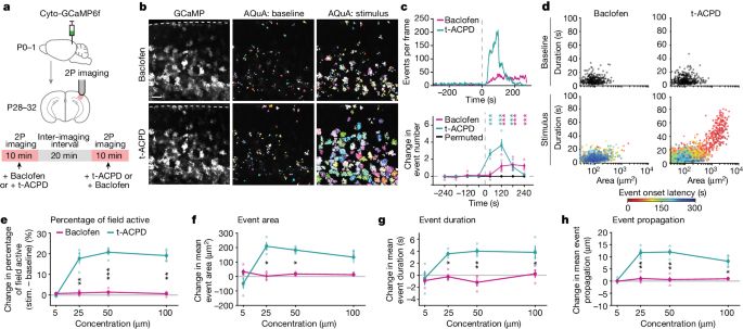
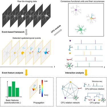
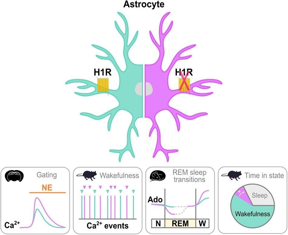
Network-level encoding of local neurotransmitters in cortical astrocytes - Nature
A study investigates subcellular, single-cell and network-level comunication within the astrocyte network in response to the two major neurotransmitter inputs.
www.nature.com