George Campbell
@geobellward.bsky.social
900 followers 570 following 120 posts
Light microscopy facility staff with interest in learning and sharing information about sample preparation, image acquisition, and image display best practices. Keen interest in Expansion Microscopy.
Posts Media Videos Starter Packs
geobellward.bsky.social
Yes, that fixed it. I can open each one and Save as LUT and back into the LUTs folder. They now show up in LUTs manager as anything else would.
geobellward.bsky.social
They don't. If I open them in Notepad, they (256 rows of 3 8-bit values) don't seem to have the same format as the other .lut files, so that must be the issue, although I can use them in all other applications. As you've made some LUTs, is there an explanation of how to format a .lut file properly?
geobellward.bsky.social
one more excellent feature: set it up to run on Fiji launch by catching the command in the macro Recorder

Edit -> Options -> Startup
run("Channels and Contrast");
🥳
geobellward.bsky.social
Hmm, I have a couple of manually added LUTs in my Fiji. Is there a way to get them to be read into the LUT manager also? They are in the luts folder and show up in the Lookup Table menu and in the NeuroCytoLUTs menu (after a few tweaks).
geobellward.bsky.social
Yes, I was wondering how the estimated description and colors were assigned (and what happens if someone had additional LUTs). Cool, thanks!
geobellward.bsky.social
one question... how is the LUTs finder annotated/organized?
geobellward.bsky.social
Very cool features so far! My favorite features : grayscale preview, color groupings, favorite LUTs, merge/split/reorder menus within easy reach, perceptual uniformity scale
geobellward.bsky.social
Can’t wait to give this a whirl on Monday!
Reposted by George Campbell
kwolbachia.bsky.social
I’m happy to share some plugins I’ve been developping this summer: "Channels and Contrast" and LUTs Manager!
I can’t find new bugs and ideas by now so I need your help to please test them in your machines and report bugs, feedbacks and ideas! forum.image.sc/t/looking-fo...
Reposted by George Campbell
biovoxxel.de
Bio-Imaging with Shakespeare: pseudocoloring (LUTs) — reveal hidden imaging issues and highlight important data.

✔ Spot noise, shading & saturation
✔ Map data for spatial insight
✔ Make color accessible to all
✔ Why to use a calibration bar

Full guide, tools & tutorial inside 👇
buff.ly/8x7Shiq
geobellward.bsky.social
The Zeiss 63X/1.15 WI 0.6mm WD is our workhorse right now.
Reposted by George Campbell
ritastrack.bsky.social
#GEF25 the expansion microscopy community would benefit from high NA, long working distance water objectives. Who's working on these? What's out there?
Reposted by George Campbell
nadjahuempf.bsky.social
Now out on bioRxiv. 🥳My research on #cytokinesis, averaging thousands of #ExM images🔬, creating a dynamic atlas of cytokinesis 🦠⏳. Here's an animated sneak peek of what we found. Better resolution on bioRxiv😄 #PSFoftheGIF
Reposted by George Campbell
christlet.bsky.social
#FluorescenceFriday: getting ready for #GEF25, playing with Ciarán's stunning ExM data
geobellward.bsky.social
Awesome, I am impressed with the flatness of the bottom surface of the cell. I see curvature of the bottom surface in some my samples, and I'm trying to identify the cause. Looks like I need to try some U-ExM!
geobellward.bsky.social
Animated z-stack of fixed and expanded cell(s). Perhaps I should add the height per frame to clarify.
geobellward.bsky.social
I am using a TREx-1000 protocol (for ~4X expansion via 1000ug/mL bis-acrylamide) with a few small adjustments.

The actin stain is pre-gelation staining via Chrometra's modified phallodin probes (based on the TRITON probes from the Hofkens lab)
geobellward.bsky.social
Thank you for correcting me, I truly appreciate it and wish that I could edit the post. Although mitosis is not my background (obviously I suppose!), I always find mitosis amazing and stop to image it. I will be more careful with my descriptions in the future.
geobellward.bsky.social
I am also very curious. The cell looks to be in wonderful shape!
geobellward.bsky.social
Meanwhile, I'll add the info here:

Fibroblasts prepared for ExM and imaged on a spinning disk confocal microscope.
DNA in purple (BIOP-ElectricIndigo)
Outer mitochondrial membrane in blue (BIOP-Azure)
F-actin in grayscale
alpha-tubulin in orange (BIOP-Amber)
LUTs selected via NeuroCytoLUTs plugin
geobellward.bsky.social
Actually, I don't see how to read the alt-text on a video. Anyone know?
geobellward.bsky.social
Bringing some more ExM to #FluorescenceFriday with what appears to be a frustrated cell division in fibroblasts.

More details in Alt-text.
Reposted by George Campbell
nicolasdenans.bsky.social
The WB-ExM protocol described here works with.... every sample we tested! Here a 3do quail embryo (white= pan-protein; red=MF20) www.biorxiv.org/content/10.1...
Reposted by George Campbell
bethcimini.bsky.social
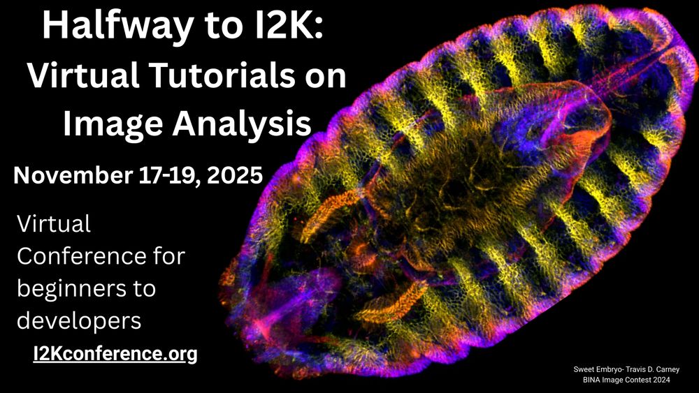
Halfway to I2K is BACK, friends of all kinds! Last year, 650 people attended 30+ TOTALLY FREE image analysis workshops of all kinds, across many timezones.

If you make image analysis software and want to teach it, workshop submissions are open now! We'd love to have your tool highlighted.
bioimagingna.bsky.social
#HappyFluorescenceFriday!

#microscopycommunity- want to learn open source image analysis or share your knowledge to help others? We’ve got a FREE virtual workshop Nov 17-19! Now accepting workshop session applications!

Learn more & sign up: buff.ly/esGIotD
geobellward.bsky.social
Amazing view, and so Talley. Jealous of those shoes but watch your ankles!