Hans Clevers
@hansclevers.bsky.social
1.2K followers 22 following 30 posts
-Scientist interested in Wnt signaling, stem cells and organoids -Director of pRED/Roche's Institute of Human Biology Basel CH. -Professor and group leader in Utrecht at the Hubrecht Institute and the Princess Maxima Center
Posts Media Videos Starter Packs
hansclevers.bsky.social
thecleverslab.bsky.social
4/4 This work was a true team effort between @amolf-nl.bsky.social, IHB Basel, and the @Hubrechtinstitute.bsky.social — led by @danielkrueger.bsky.social and Kasper Spoelstra. Huge thanks to all collaborators for making it possible.
hansclevers.bsky.social
11/11Gut homeostasis is not passive crowd control. It’s an active, force-regulated process. Cells constantly test each other’s strength; those that fail the tug-of-war extrude.A collaboration between the @hubrechtinstitute.bsky.social , @amolf-nl.bsky.social & @IHB lead by @danielkrueger.bsky.social
hansclevers.bsky.social
10/11 Disease link: In EpCAM loss (tufting enteropathy), hyperactive myosin caused excessive extrusion, growth defects, and tissue disruption. Too much force destabilizes the barrier. Mixing mutant & WT cells made healthy cells extrude. Myosin inhibition rescued this effect.
hansclevers.bsky.social
9/11 With point ablations at the base, we intentionally weakened individual cells. Within minutes, these cells extruded - loss of tension likely triggers extrusion.
hansclevers.bsky.social
8/11 Local optogenetic activation of cells on villus substrates, creating boundaries between highly- and less-contractile cells caused preferential extrusion on the weaker side. In mosaic organoids, less contractile cells were consistently eliminated.
"Weaker" less contractile cells that can not sustain the tension level of their neighbours in the tissue preferentially extruded.
hansclevers.bsky.social
7/11 Optogenetics let us induce contractility with blue light and spatial precision. Extrusion rates jumped up upon activation, showing that globally increased tension directly drives extrusion.
hansclevers.bsky.social
6/11 A basal actomyosin network drives epithelial tension. On hydrogel villus substrates (generated @ IHB, Basel), resembling the in vivo topology, we saw pulsatile contractions—one cell contracts, neighbors expand. A tissue-wide tug-of-war.
hansclevers.bsky.social
5/11 Using CRISPR-generated myosin reporters, we first observed myosin increasing in extruding cells, together with a synchronized response in their neighbors—explaining extrusion dynamics as a coordinated process.
hansclevers.bsky.social
4/11 Are villus tips—where extrusion peaks—compressed by crowding? At the Hubrecht, laser ablation revealed they are under tension, not compression. That tension sets the stage for extrusion.
Laser ablation at the villus tip revealed that the tissue is under tension and cells are not under compression.
hansclevers.bsky.social
3/11 Using long-term live imaging & tracking (with @amolf-nl.bsky.social), we followed hundreds of extrusion events. Most villus cells extruded alive, not apoptotic. Extrusion did not correlate with density.
hansclevers.bsky.social
2/11 The intestinal lining renews every few days. Old cells are shed by extrusion—a process essential for barrier integrity and linked to diseases like IBD. But what actually triggers extrusion in mammals was unclear.
hansclevers.bsky.social
1/11 In @science.org: A new perspective on how our intestines renew. Cells are not “pushed out” by crowding or die from apoptosis. Instead, cells play a mechanical tug-of-war, where weaker cells extrude, reframing gut renewal as force-regulated. www.science.org/doi/10.1126/...
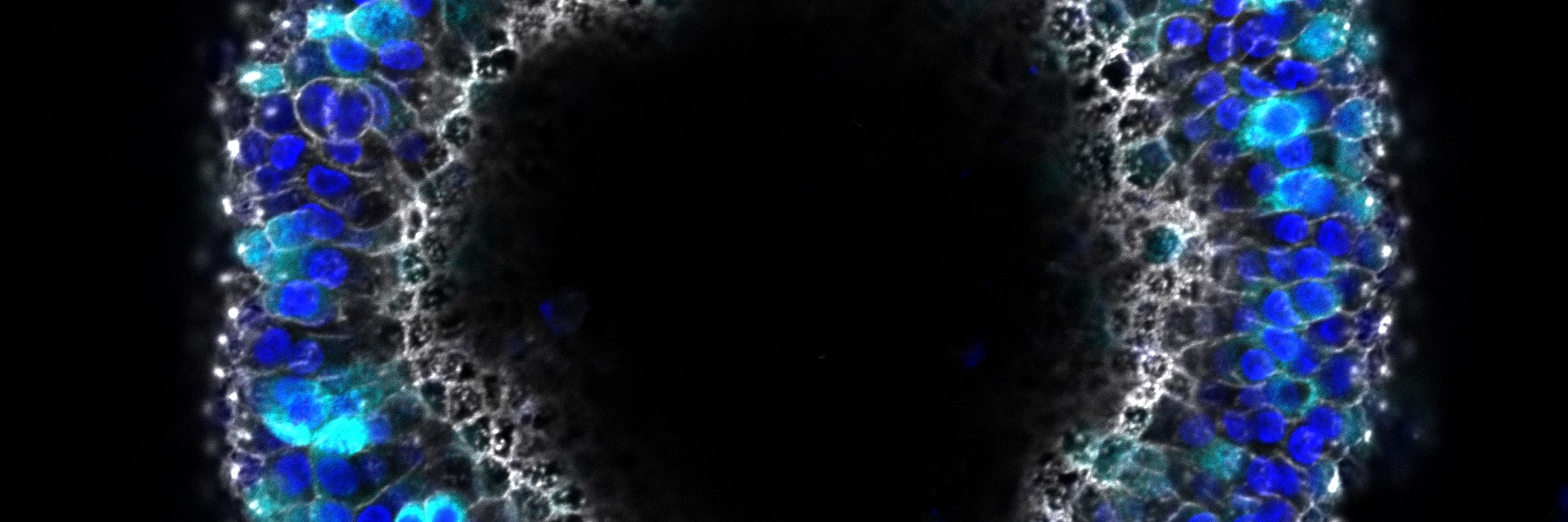
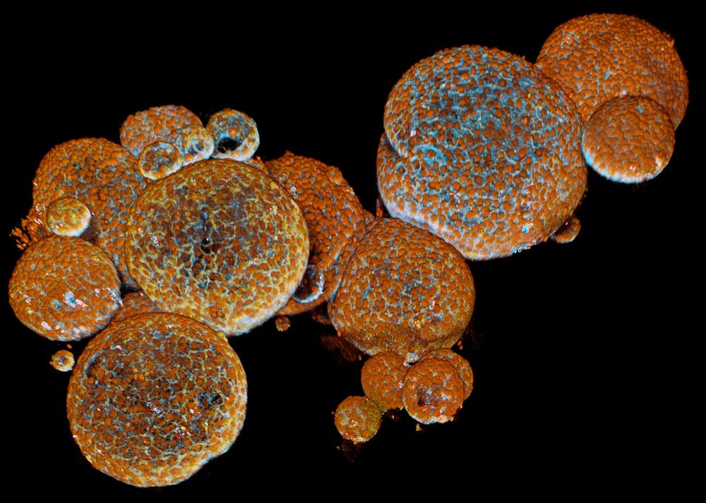
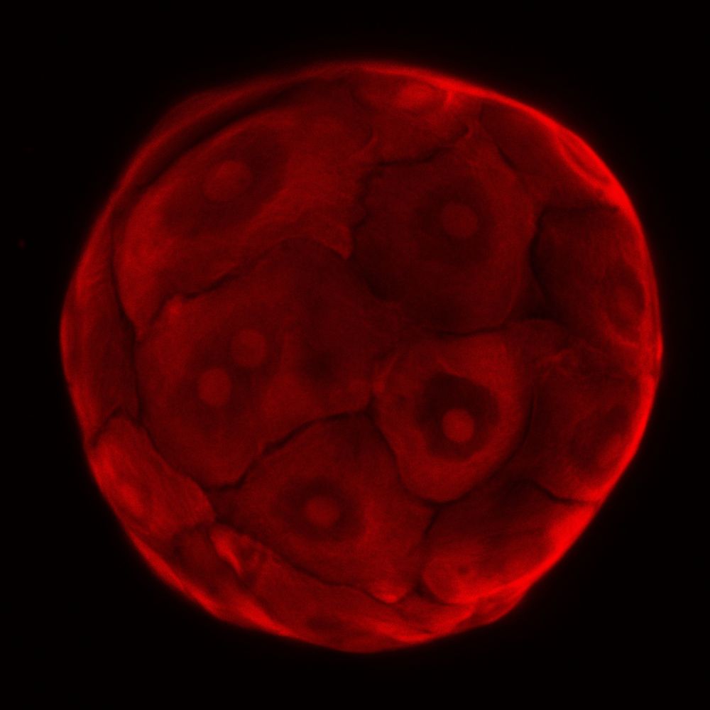
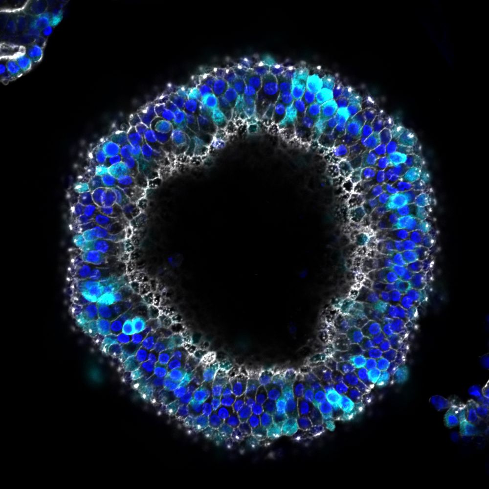
Epithelial tension controls intestinal cell extrusion
Cell extrusion is essential for homeostatic self-renewal of the intestinal epithelium. Extrusion is thought to be triggered by crowding-induced compression of cells at the intestinal villus tip. In th...
www.science.org
hansclevers.bsky.social
Brain diseases are tough to treat because of the blood-brain barrier. For decades, it stumped drug delivery. At Roche pRED, we challenged that! Our Brainshuttle™ technology safely transports drugs across, opening new doors for CNS therapies. Learn more: www.roche.com/stories/clev...
Roche | Clever delivery
Brainshuttle™ technology by Roche enables effective medicine delivery across the blood-brain barrier, revolutionising CNS disease treatment and patient care.
www.roche.com
hansclevers.bsky.social
Exciting #organoids meeting in Glasgow, just after the summer!
hansclevers.bsky.social
🔬 Developing next-generation Antibiotics: @Roche we are pioneering the discovery and development of novel antibiotics that can fight resistant bacteria, aiming for future generations to have effective treatments. Visit our #AMR hub:https://www.roche.com/stories/antibiotics
hansclevers.bsky.social
@NatureMedicine
invited me to reflect on leading research in academia vs. industry. In the end, all -academia, biotech & pharma- is needed to translate scientific breakthroughs into scalable treatments. rdcu.be/elPt8
@_hubrecht
@Roche
Parallel worlds in pharma and academia
Nature Medicine - Pharma and academia require different ways of working and speaking, but teamwork always wins.
rdcu.be
hansclevers.bsky.social
Snake venom gland organoids. Study by Yorick Post and collaborators. www.sciencedirect.com/science/arti...
Image credit: Anne Rios
hansclevers.bsky.social
a human liver organoid
hansclevers.bsky.social
#Lgr5+ gut #stemcells don't divide asymmetrically. They simple divide and then compete for space: This is called neutral competition. Stem cell numbers are set by the available space between the #Panethcells. Paper: Hugo Snippert et al. Cell (2010). www.cell.com/action/showP...
hansclevers.bsky.social
📢Roche's Institute of Human Biology seeks 2 visionary scientific leaders: Head of Computational Biology & Head of Exploratory Biology. Drive pioneering research, shape scientific strategy and lead innovation at the crossroad of academia and industry!🔗https://go.roche.com/ls0un #organoids #TeamIHB
Image credit: Silvia Berger, Saba Rezakhani, Irineja Cubela (Roche's IHB) 
About the image: Kidney organoids with glomeruli - paraffin section stained for nuclei (blue). Glomerular markers: Nephrin (red) and Synaptopodin (green), and tubule marker EpCam (grey). Acquired with Nikon Ti2 25x water immersion objective.
Reposted by Hans Clevers
jimjansen.bsky.social
Ik was in Basel voor een interview met @hansclevers.bsky.social

“Het is een proces van eindeloze een-tweetjes. En bij de start van de eerste fase van het klinisch onderzoek moet je al in je hoofd hebben wat voor patiëntengroep je over zes jaar wilt behandelen.”

www.newscientist.nl/blogs/een-me...
‘Een medicijn ontwikkelen is een proces van eindeloze een-tweetjes’
‘Medicijnen ontwikkelen bij Roche is schaken in een matrix van twaalf dimensies’, zegt Hans Clevers, hoogleraar moleculaire genetica.
www.newscientist.nl
hansclevers.bsky.social
3D Reconstruction of human fetal pancreatic organoid with an endogenous (knock-in) reporter for the endocrine cell marker Chromogranin-A (green), while stained for the acinar cell marker CarboxyPeptidase A1/2 (magenta) and nuclei, DAPI (blue)
hansclevers.bsky.social
Clonal organoids grown from a single LGR5+ cell recapitulate this tri-potency in vitro. Our study describes a human fetal tri-potent stem cell, capable of long-term expansion in vitro and of generating all three pancreatic cell lineages 5/
hansclevers.bsky.social
The four lines expand exponentially for >2 years under optimized culture conditions. Single cell RNA seq identifies rare LGR5+ cells in fetal pancreas and in hfPOs as the root of the developmental hierarchy. These LGR5+ cells share multiple markers with adult gut stem cells 4/
hansclevers.bsky.social
Mouse studies indicate that these three compartments derive from a transient, common pancreatic progenitor. Amanda et al derive 18 human fetal pancreas organoid (hfPO) lines from gestational week 8-17 samples. Four lines generate acinar-, ductal- and endocrine lineage cells 3/