Jana Kroll
@janakroll.bsky.social
190 followers
240 following
20 posts
Postdoc at MDC Berlin
cryo-electron tomography, neuronal synaptic transmission, optogenetics
Posts
Media
Videos
Starter Packs
Pinned

Reposted by Jana Kroll

Reposted by Jana Kroll
Reposted by Jana Kroll

Reposted by Jana Kroll


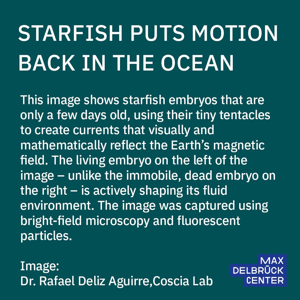
Jana Kroll
@janakroll.bsky.social
· May 13
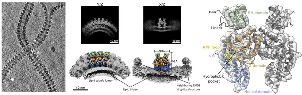
Jana Kroll
@janakroll.bsky.social
· May 12


Reposted by Jana Kroll
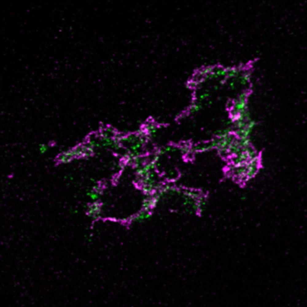

Jana Kroll
@janakroll.bsky.social
· Feb 16
Jana Kroll
@janakroll.bsky.social
· Feb 15
Jana Kroll
@janakroll.bsky.social
· Feb 14
Jana Kroll
@janakroll.bsky.social
· Feb 13
Jana Kroll
@janakroll.bsky.social
· Feb 13


Jana Kroll
@janakroll.bsky.social
· Feb 12