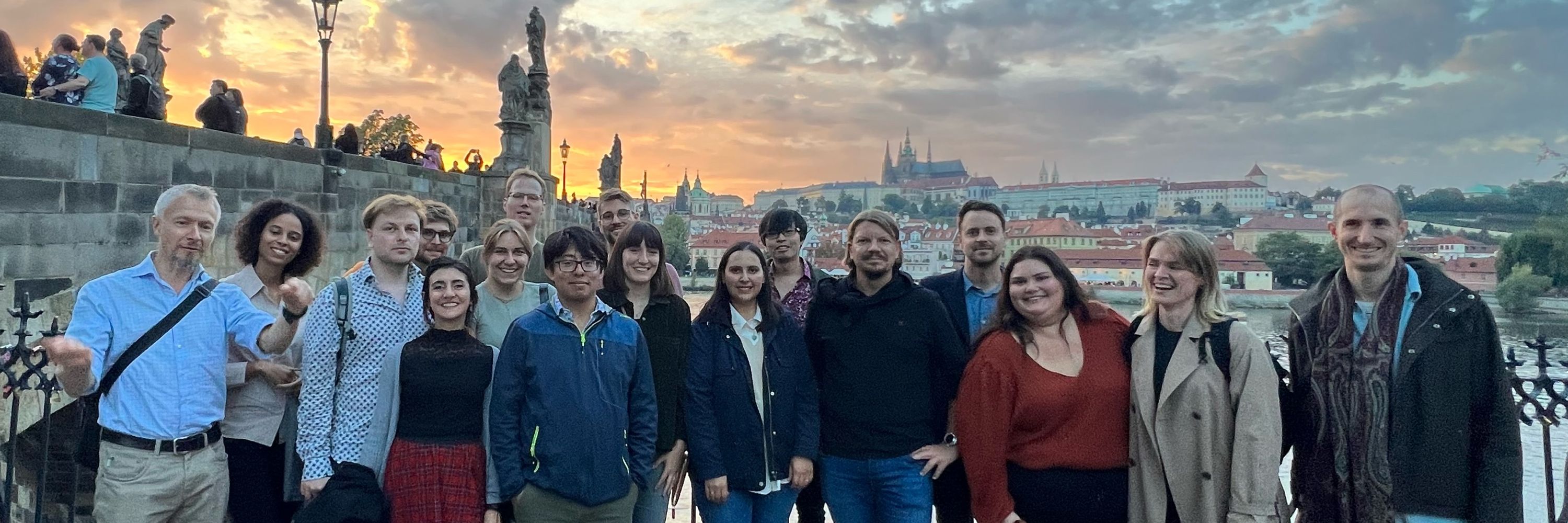
JesperOlsenLab
@jesperolsenlab.bsky.social
630 followers 90 following 36 posts
Mass spectrometry for quantitative proteomics. The Olsen group at the Center for Protein Research, University of Copenhagen.
Posts Media Videos Starter Packs
jesperolsenlab.bsky.social
We made a short video for the ERA PerMed video competition 🎬
We present our collaborative project on “personalising clinical decisions in ovarian cancer through patient-derived in vitro models”.
Have a look at it 👇🏼 and don’t forget to suport us by putting like on Youtube 👍🏼

youtu.be/J4bc1MPqIqE?...
OVA-PDM - Personalising clinical decisions in ovarian cancer through patient-derived in vitro models
YouTube video by European Partnership for Personalised Medicine
youtu.be
jesperolsenlab.bsky.social
With a 40% reduction in analysis time and >12,000 proteins covered in 2.7 hours, it's setting new benchmarks for proteome studies.
jesperolsenlab.bsky.social
The new Astral Zoom MS overcomes previous MS limitations and is now achieving ultra-fast scans up to 270 Hz. It allows to identity > 100,000 peptides with short LC gradients.
jesperolsenlab.bsky.social
The combination of pre accumulation and faster scans significantly improves peptide/protein IDs on short LC gradients and boosts sensitivity for high-throughput workflows 📈
The benefits are even higher with ΦSDM, especially in fast, low-resolution Orbitrap runs.
jesperolsenlab.bsky.social
Florian Harking & Ulises H. Guzman made it happen by preaccumulating ions in the bent flatapole and applying phase-constrained spectrum deconvolution method (ΦSDM).
jesperolsenlab.bsky.social
🦷 Excited to share how proteomics is pushing the boundaries of palaeoanthropology!
We used proteomics on Paranthropus robustus fossils from South Africa (~2 million years old) to get insights into biological sex and variation.
A great example of how LC-MS/MS can uncover new transformative info👇
palaeoprotpalesa.bsky.social
Hello,

Our paper on enamel proteins from Paranthropus robustus has finally been peer reviewed, please have a read here: www.science.org/doi/10.1126/...

Paranthropus robustus has been puzzling scientists since its discovery in 1938 in South Africa, where a high number of fossils have been found.
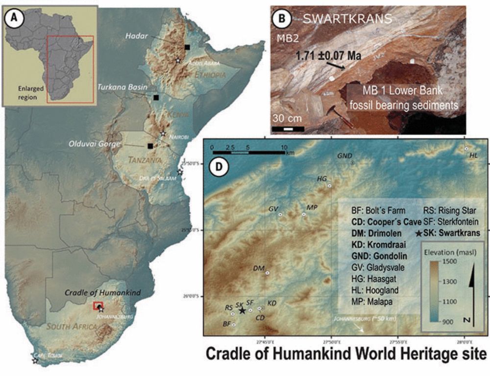
Enamel proteins reveal biological sex and genetic variability in southern African Paranthropus
Paranthropus robustus is a morphologically well-documented Early Pleistocene hominin species from southern Africa with no genetic evidence reported so far. In this work, we describe the mass spectrome...
doi.org
jesperolsenlab.bsky.social
Knocking down CAVIN1 in a 3D model reveals its connection with tumour invasiveness. Hybrid-DIA based phosphoproteomics profiling reveals an intriguing link between mTOR-signalling and its potential association to CAVIN1.
jesperolsenlab.bsky.social
The mesenchymal-like subtype was shown to be marked by increased CAVIN1 and PRELP. Higher CAVIN1 correlates with higher relapse risk in two independent cohorts. We confirmed this in a second cohort and were able to link this to Epithelial to Mesenchymal Transition.
jesperolsenlab.bsky.social
We stratified 412 CRC tumors into 4 distinct subtypes using quantitative proteomics. Our results show clear links to transcriptomics CMS, immune infiltration, tumor purity, and relapse.
jesperolsenlab.bsky.social
We also identified aberrant PDGF receptor (PDGFR) activation as a key feature of OCSCs.
When we treated patient-derived ovarian cancer cells with a PDGFR inhibitor, their stemness potential dropped (sphere formation ↓).
jesperolsenlab.bsky.social
Our stemness signature correlates with chemo-refractoriness in clinical proteomics data.
That’s huge: it means this signature might help predict therapy resistance.
jesperolsenlab.bsky.social
Stemness-associated proteins were involved in extracellular matrix remodeling.
This points to a link between the stem-like state and the tumor microenvironment.
jesperolsenlab.bsky.social
To validate our stemness signature, we used single-sample GSEA on published proteomics data from embryonic stem cell differentiation.
jesperolsenlab.bsky.social
We profiled the proteome and phosphoproteome of 3D spheres enriched for OCSCs from 10 treatment-naive patient-derived lines.
This revealed a shared stemness-related signature across patients, despite high inter-patient heterogeneity.
jesperolsenlab.bsky.social
We confirmed this observation and exploited it to compare different modalities of cell division arrest in human foreskin fibroblasts.
jesperolsenlab.bsky.social
Histone turnover enables distinguishing dividing cells from slow/non-dividing ones, due to the extremely slow core histone turnover in non-dividing cells.
jesperolsenlab.bsky.social
Protein turnover provides an orthogonal measurement to protein abundance, revealing functional differences between cell types and states, and improving protein co-regulation analysis to determine members of protein complexes.
jesperolsenlab.bsky.social
We then performed a large-scale SC-pSILAC experiment to study the undirected differentiation of human induced pluripotent stem cells through embryoid body induction in >1000 individual single cells.
Our comprehensive dataset shows protein turnover dynamics during cell differentiation over 2 months.
jesperolsenlab.bsky.social
We first studied treatments altering global protein turnover including proteasome(bortezomib), protein translation (cycloheximide), and beta-cathenin inhibitions for altered turnover of a specific protein(GSK3 inhibitor).
SC-pSILAC revealed treatment-related changes in single cells.
jesperolsenlab.bsky.social
🚀 Excited to share our latest article in #singlecell proteomics published in Cell!
We've developed SC-pSILAC to simultaneously measure protein turnover and abundance in single cells, unlocking the first large-scale, 2D proteomic insights at single-cell resolution!

www.cell.com/cell/fulltex...
Global analysis of protein turnover dynamics in single cells
The SC-pSILAC method enables single-cell measurement of both protein abundance and turnover, providing notable advances in the depth and versatility of proteomic technologies.
eur02.safelinks.protection.outlook.com
jesperolsenlab.bsky.social
Finally fixed cells showed altered morphology vs. non fixed cells. Yet, after freezing the morphological changes in fixed cells are reduced compared to non fixed cells. Fixed frozen single cells have significantly lower CVs than non fixed frozen cells, enhancing data quality and reproducibility.