Fernando Santana
@lfsantana68.bsky.social
110 followers
120 following
24 posts
🔬Physiologist & Biophysicist | Vice Dean for Basic Sciences and Chair of Physiology & Membrane Biology
🧠 Champion of thematic breadth in science & medical research
📢 Exploring how structure fuels innovation
☕ Fueling ideas with coffee & conversation
Posts
Media
Videos
Starter Packs

Reposted by Fernando Santana


Fernando Santana
@lfsantana68.bsky.social
· Aug 22

Beat–locked ATP microdomains in the sinoatrial node map a calcium–timed energetic hierarchy and regional pacemaker roles
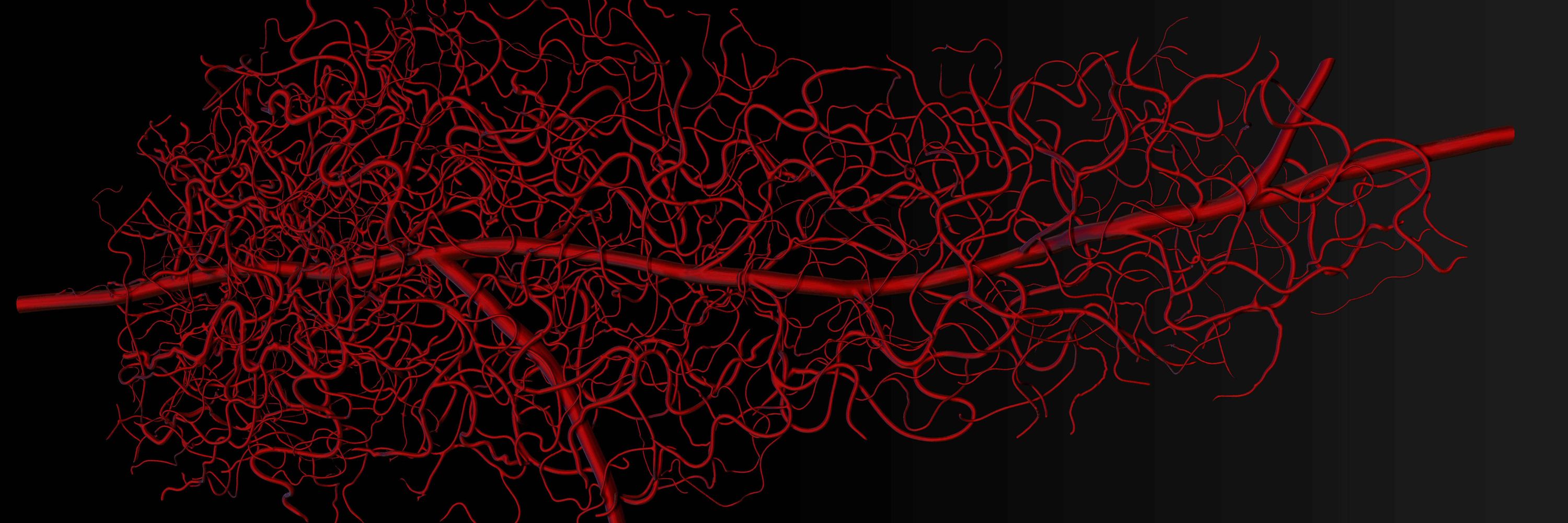
Pacemaker myocytes of the sinoatrial (SA) node initiate each heartbeat through coupled voltage and Ca2+ oscillators, but whether ATP supply is regulated on a beat–by–beat schedule in these cells has been unclear. Using genetically encoded sensors targeted to the cytosol and mitochondria, we tracked beat–resolved ATP dynamics in intact mouse SA node and isolated myocytes. Cytosolic ATP rose transiently with each Ca2+ transient and segregated into high– and low–gain phenotypes defined by the Ca2+–ATP coupling slope. Mitochondrial ATP flux adopted two stereotyped waveforms—Mode–1 ″gains″ and Mode–2 ″dips″. Within Mode–1 cells, ATP gains mirrored the cytosolic high/low–gain dichotomy; Mode–2 dips scaled linearly with Ca2+ load and predominated in slower-firing cells. In the intact node, high–gain/Mode–1 phenotypes localized to superior regions and low–gain/Mode–2 to inferior regions, paralleling gradients in rate, mitochondrial volume, and capillary density. Pharmacology placed the Ca2+ clock upstream of ATP production: the HCN channel blocker ivabradine slowed the ATP cycle without changing amplitude, whereas the SERCA pump inhibitor thapsigargin or the mitochondrial uncoupler FCCP abolished transients. Mode–2 recovery kinetics indicate slower ATP replenishment that would favor low–frequency, fluctuation–rich firing in a subset of cells. Together, these findings reveal beat-locked metabolic microdomains in which the Ca2+ clock times oxidative phosphorylation under a local O2 ceiling, unifying vascular architecture, mitochondrial organization, and Ca2+ signaling to coordinate energy supply with excitability. This energetic hierarchy helps explain why some SA node myocytes are more likely to set rate whereas others may widen bandwidth. ### Competing Interest Statement The authors have declared no competing interest. National Heart Lung and Blood Institute, https://ror.org/012pb6c26, HL168874 American Heart Association, https://doi.org/10.58275/AHA.25POST1378853.pc.gr.227467
www.biorxiv.org
Reposted by Fernando Santana

Manuel F. Navedo
@mfnavedo.bsky.social
· Aug 15

Arterial Myocyte Pannexin 1 Channel Controls Vascular Reactivity in Diabetic Hyperglycemia | Circulation Research
BACKGROUND: Diabetic hyperglycemia promotes vasoconstriction by activating an ATP-dependent P2Y11L (P2Y11-like receptor)/AC5 (adenylyl cyclase 5)/AKAP5 (A-kinase anchoring protein 5)/PKA
(protein kina...
www.ahajournals.org
Fernando Santana
@lfsantana68.bsky.social
· Aug 13


Reposted by Fernando Santana

Reposted by Fernando Santana

Jeremy Berg
@jeremymberg.bsky.social
· Jul 31
Fernando Santana
@lfsantana68.bsky.social
· Jul 28
Fernando Santana
@lfsantana68.bsky.social
· Jul 27
Reposted by Fernando Santana


Fernando Santana
@lfsantana68.bsky.social
· Jul 23
Reposted by Fernando Santana


Fernando Santana
@lfsantana68.bsky.social
· Jul 11
Demonstration of Beat-to-Beat, On-Demand ATP Synthesis in Ventricular Myocytes Reveals Sex-Specific Mitochondrial and Cytosolic Dynamics.
The substantial energetic demands on ventricular myocytes imposed by the transport of ions and cross–bridge cycling associated with each heartbeat are well known, yet the spatiotemporal dynamics of AT...
www.biorxiv.org
Reposted by Fernando Santana
Manuel F. Navedo
@mfnavedo.bsky.social
· Jul 10

Reposted by Fernando Santana


Reposted by Fernando Santana


Reposted by Fernando Santana