


LifeCanvas Technologies
@lifecanvastech.bsky.social
Pioneering end-to-end solutions for 3D spatial biology.
www.lifecanvastech.com
www.lifecanvastech.com
You've seen our transparent samples... now we want to see YOURS! If you're sharing 3D #MicroscopyMonday data, we know you've got some forbidden gummies floating around the lab.
Post your cleared tissues, tag us, and snag a discount for your next order of SmartBatch+ reagent kits! #tissueclearing
Post your cleared tissues, tag us, and snag a discount for your next order of SmartBatch+ reagent kits! #tissueclearing

December 8, 2025 at 3:46 PM
You've seen our transparent samples... now we want to see YOURS! If you're sharing 3D #MicroscopyMonday data, we know you've got some forbidden gummies floating around the lab.
Post your cleared tissues, tag us, and snag a discount for your next order of SmartBatch+ reagent kits! #tissueclearing
Post your cleared tissues, tag us, and snag a discount for your next order of SmartBatch+ reagent kits! #tissueclearing
Change how you see 👀 your samples with #DALISPIM light-sheet imaging. Here, nuclear staining (magenta) shows thick layers of retinal cells on the back of the eyeball connecting to the optic nerve, with a thin layer of cells over the cornea. What else do you want to see labeled? #FluorescenceFriday
December 5, 2025 at 3:20 PM
Change how you see 👀 your samples with #DALISPIM light-sheet imaging. Here, nuclear staining (magenta) shows thick layers of retinal cells on the back of the eyeball connecting to the optic nerve, with a thin layer of cells over the cornea. What else do you want to see labeled? #FluorescenceFriday
We’re gearing up to head to #CellBio2025, the joint annual meeting of @ascbiology.bsky.social and @embo.org. Meet up with our team at booth 228 to learn how whole-tissue imaging can add a new dimension to your #cellbiology research. 🧫
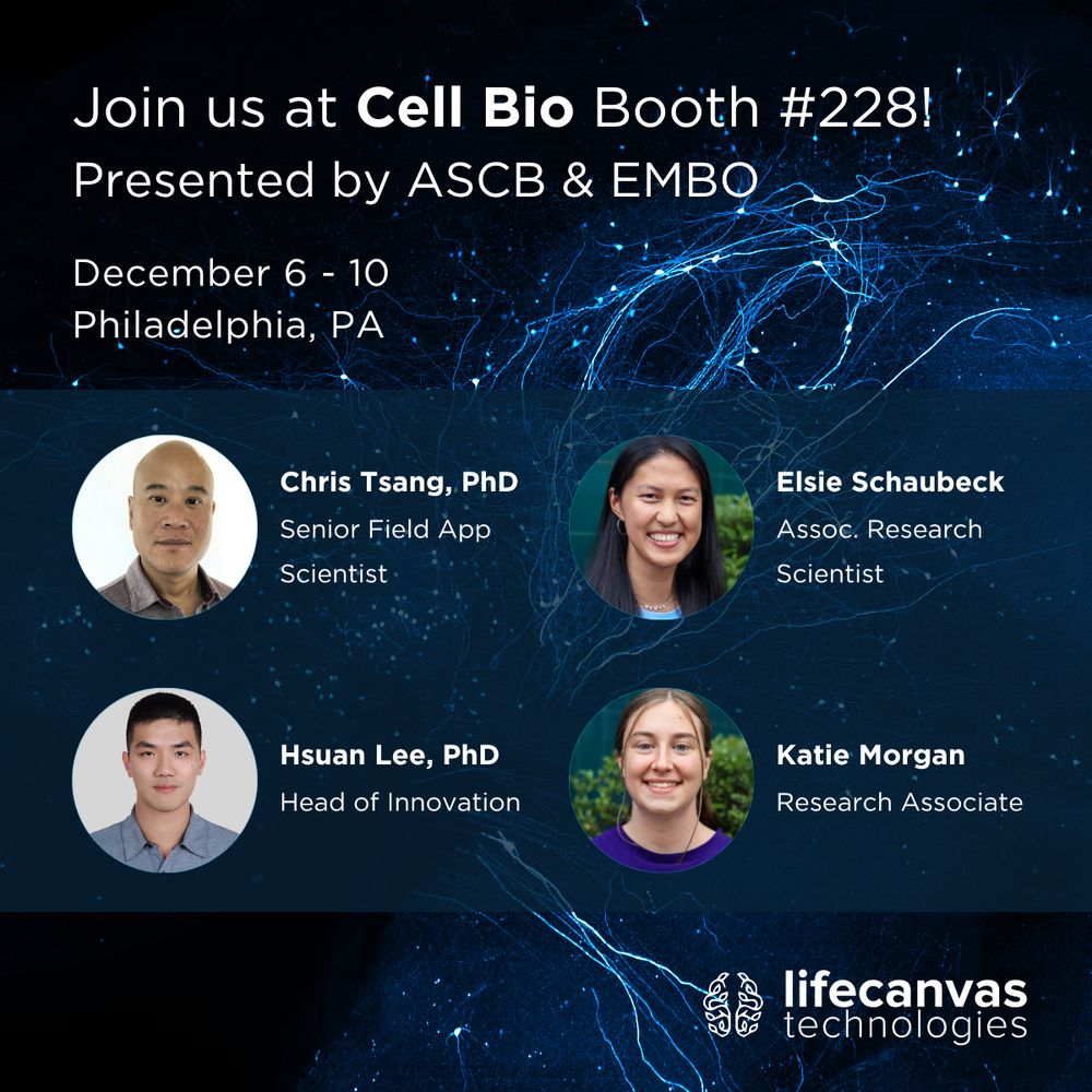
December 4, 2025 at 3:18 PM
We’re gearing up to head to #CellBio2025, the joint annual meeting of @ascbiology.bsky.social and @embo.org. Meet up with our team at booth 228 to learn how whole-tissue imaging can add a new dimension to your #cellbiology research. 🧫
What can you discover with one powerful, streamlined pipeline for 3D #SpatialBiology?
Researchers in @katrinachoe.bsky.social's lab used whole-brain labeling, clearing, imaging, and ML-based cell detection to quantify OXT+ neurons across part of the hypothalamus called the PVN 🧵 (1/4)
Researchers in @katrinachoe.bsky.social's lab used whole-brain labeling, clearing, imaging, and ML-based cell detection to quantify OXT+ neurons across part of the hypothalamus called the PVN 🧵 (1/4)

December 3, 2025 at 3:47 PM
What can you discover with one powerful, streamlined pipeline for 3D #SpatialBiology?
Researchers in @katrinachoe.bsky.social's lab used whole-brain labeling, clearing, imaging, and ML-based cell detection to quantify OXT+ neurons across part of the hypothalamus called the PVN 🧵 (1/4)
Researchers in @katrinachoe.bsky.social's lab used whole-brain labeling, clearing, imaging, and ML-based cell detection to quantify OXT+ neurons across part of the hypothalamus called the PVN 🧵 (1/4)
Our data engineering & analysis team is excited to trade New England winter for sunny San Diego to attend #NeurIPS2025! We're especially looking forward to the workshop on "Imageomics: Discovering Biological Knowledge from Images Using AI". @neuripsconf.bsky.social #bioimaging

December 1, 2025 at 3:07 PM
Our data engineering & analysis team is excited to trade New England winter for sunny San Diego to attend #NeurIPS2025! We're especially looking forward to the workshop on "Imageomics: Discovering Biological Knowledge from Images Using AI". @neuripsconf.bsky.social #bioimaging
A new uniQure study shows promise for improving AAV-based #genetherapy delivery to the CNS. The team used the LifeCanvas pipeline to visualize biodistribution throughout whole mouse brains. 🐭 🧠 Learn more & see their #SmartSPIM data: bit.ly/4rixY3v

November 25, 2025 at 4:35 PM
A new uniQure study shows promise for improving AAV-based #genetherapy delivery to the CNS. The team used the LifeCanvas pipeline to visualize biodistribution throughout whole mouse brains. 🐭 🧠 Learn more & see their #SmartSPIM data: bit.ly/4rixY3v
Happy #MicroscopyMonday! Our tools allow #neuroscience researchers like Dr. Jing Zhou (Pleasure Lab, UCSF) to visualize neuronal projections with stunning detail. This video highlights the intricate wiring of cortical excitatory neurons (red) in an Emx1-Cre; Ai14 mouse brain.
November 24, 2025 at 2:56 PM
Happy #MicroscopyMonday! Our tools allow #neuroscience researchers like Dr. Jing Zhou (Pleasure Lab, UCSF) to visualize neuronal projections with stunning detail. This video highlights the intricate wiring of cortical excitatory neurons (red) in an Emx1-Cre; Ai14 mouse brain.
Aggrecan is a key component of perineuronal nets (PNNs), extracellular structures that surround certain neuronal sub-populations and regulate neuroplasticity. This video shows aggrecan in an intact spinal cord, processed & imaged with our pipeline. #FluorescenceFriday
November 21, 2025 at 2:47 PM
Aggrecan is a key component of perineuronal nets (PNNs), extracellular structures that surround certain neuronal sub-populations and regulate neuroplasticity. This video shows aggrecan in an intact spinal cord, processed & imaged with our pipeline. #FluorescenceFriday
At #SfN25 this year, we had the opportunity to support both the American Society for Chinese Neuroscientists (ASCN) and the Association of Korean Neuroscientists (AKN). We had a wonderful time connecting with researchers at both social events over food, games, and talks!

November 20, 2025 at 4:11 PM
At #SfN25 this year, we had the opportunity to support both the American Society for Chinese Neuroscientists (ASCN) and the Association of Korean Neuroscientists (AKN). We had a wonderful time connecting with researchers at both social events over food, games, and talks!
The New England biologist impulse to do science on a tick you pulled off of yourself – this is ours from this summer!
November 19, 2025 at 9:24 PM
The New England biologist impulse to do science on a tick you pulled off of yourself – this is ours from this summer!
In a global effort to map #neurodevelopment across species, scientists in the BRAIN Initiative Cell Atlas Network (BICAN) used single-cell & spatial -omics approaches combined with imaging tools like #SmartSPIM to create detailed cell-type brain atlases: nature.com/collections/... @nature.com

November 19, 2025 at 2:15 PM
In a global effort to map #neurodevelopment across species, scientists in the BRAIN Initiative Cell Atlas Network (BICAN) used single-cell & spatial -omics approaches combined with imaging tools like #SmartSPIM to create detailed cell-type brain atlases: nature.com/collections/... @nature.com
Dr. Jacquelyn Salzbank et al. @columbiamed.bsky.social used LifeCanvas whole 🧠 visualization of #microglia to investigate the role of a placental hormone in sex-specific neurodevelopmental dysfunction. Check out the paper to learn more: bit.ly/47YsH8w

November 18, 2025 at 3:14 PM
Dr. Jacquelyn Salzbank et al. @columbiamed.bsky.social used LifeCanvas whole 🧠 visualization of #microglia to investigate the role of a placental hormone in sex-specific neurodevelopmental dysfunction. Check out the paper to learn more: bit.ly/47YsH8w
Neutrophil Extracellular Traps (NETs) are fibrous structures released by immune cells to bind and kill pathogens. In collaboration with Dr. Mark Looney's lab at @ucsfhealth.bsky.social, we used #DALISPIM to visualize 3D NET formation (magenta) triggered by inflammation in a mouse lung. 🫁
November 17, 2025 at 7:51 PM
Neutrophil Extracellular Traps (NETs) are fibrous structures released by immune cells to bind and kill pathogens. In collaboration with Dr. Mark Looney's lab at @ucsfhealth.bsky.social, we used #DALISPIM to visualize 3D NET formation (magenta) triggered by inflammation in a mouse lung. 🫁
Congratulations to our SfN Travel Award winner, Dr. Élora Midavaine! Her presentation focuses on neurobiological adaptations to chronic pain during pregnancy, using LifeCanvas whole 🧠 clearing and imaging to hone in on an endogenous opioid circuit that contributes to pregnancy-induced analgesia.

November 17, 2025 at 4:38 PM
Congratulations to our SfN Travel Award winner, Dr. Élora Midavaine! Her presentation focuses on neurobiological adaptations to chronic pain during pregnancy, using LifeCanvas whole 🧠 clearing and imaging to hone in on an endogenous opioid circuit that contributes to pregnancy-induced analgesia.
Save room in your #SfN25 itinerary for some more 3D 🧠 data exploring research areas from brain development to neuroimmune pathways! Next week's poster lineup features presentations from Drs. @ingvildeb.bsky.social, Élora Midavaine, Quan Jiang, Zhen Wang, & Yidan Chen.

November 13, 2025 at 2:44 PM
Save room in your #SfN25 itinerary for some more 3D 🧠 data exploring research areas from brain development to neuroimmune pathways! Next week's poster lineup features presentations from Drs. @ingvildeb.bsky.social, Élora Midavaine, Quan Jiang, Zhen Wang, & Yidan Chen.
This weekend kicks off #SfN25! Have you made your poster game plan yet?
Today and tomorrow we'll be sharing a selection of posters featuring LifeCanvas 3D histology and imaging data 🧠 that you can add to your agenda. Check out our Sunday schedule below.
Today and tomorrow we'll be sharing a selection of posters featuring LifeCanvas 3D histology and imaging data 🧠 that you can add to your agenda. Check out our Sunday schedule below.

November 12, 2025 at 3:08 PM
This weekend kicks off #SfN25! Have you made your poster game plan yet?
Today and tomorrow we'll be sharing a selection of posters featuring LifeCanvas 3D histology and imaging data 🧠 that you can add to your agenda. Check out our Sunday schedule below.
Today and tomorrow we'll be sharing a selection of posters featuring LifeCanvas 3D histology and imaging data 🧠 that you can add to your agenda. Check out our Sunday schedule below.
In a new Nature study, Dr. @gabineiman.bsky.social et al. developed an in vitro screening method to predict transfection efficacy of LNP-mRNA complexes in the heart, validating it with #SmartSPIM imaging in whole mouse hearts. Check out the paper: bit.ly/444P0rT
@qb3-berkeley.bsky.social
@qb3-berkeley.bsky.social

November 11, 2025 at 5:50 PM
In a new Nature study, Dr. @gabineiman.bsky.social et al. developed an in vitro screening method to predict transfection efficacy of LNP-mRNA complexes in the heart, validating it with #SmartSPIM imaging in whole mouse hearts. Check out the paper: bit.ly/444P0rT
@qb3-berkeley.bsky.social
@qb3-berkeley.bsky.social
That's not discount Halloween candy on the right – it's a cleared mouse brain! We recently went down to New Orleans to visit @tulaneu.bsky.social's Brain Institute and demonstrate #tissueclearing techniques for neuroscientists. Thanks Dr. Deniz Ucar for hosting us at the Cell & Tissue Imaging Core!

November 10, 2025 at 3:42 PM
That's not discount Halloween candy on the right – it's a cleared mouse brain! We recently went down to New Orleans to visit @tulaneu.bsky.social's Brain Institute and demonstrate #tissueclearing techniques for neuroscientists. Thanks Dr. Deniz Ucar for hosting us at the Cell & Tissue Imaging Core!
📣📣 We've extended our travel award deadline to Tues, Nov 11th. A few more days left to apply for $1000 towards #SfN25 travel expenses! Just tell us how you used LifeCanvas tech in your poster. Winner announced next Friday, 11/14!

November 7, 2025 at 7:15 PM
📣📣 We've extended our travel award deadline to Tues, Nov 11th. A few more days left to apply for $1000 towards #SfN25 travel expenses! Just tell us how you used LifeCanvas tech in your poster. Winner announced next Friday, 11/14!
Glomeruli, the filtering parts of nephrons, are visible in cyan in this mouse kidney section imaged with DALISPIM. In mice, kidneys have over 10,000 nephrons!
Our new open-top #lightsheet microscope's unique design allowed us to image this 1-mm thick sample directly in a 12-well plate.
Our new open-top #lightsheet microscope's unique design allowed us to image this 1-mm thick sample directly in a 12-well plate.
November 7, 2025 at 3:57 PM
Glomeruli, the filtering parts of nephrons, are visible in cyan in this mouse kidney section imaged with DALISPIM. In mice, kidneys have over 10,000 nephrons!
Our new open-top #lightsheet microscope's unique design allowed us to image this 1-mm thick sample directly in a 12-well plate.
Our new open-top #lightsheet microscope's unique design allowed us to image this 1-mm thick sample directly in a 12-well plate.
"I have too many cool 3D microscopy images in my algorithm" - no one
Connect with us on LinkedIn and Instagram to bring some ✨fluorescence✨ into your daily scroll:
LI: www.linkedin.com/company/life...
IG: instagram.com/lifecanvas_tec
Connect with us on LinkedIn and Instagram to bring some ✨fluorescence✨ into your daily scroll:
LI: www.linkedin.com/company/life...
IG: instagram.com/lifecanvas_tec

November 6, 2025 at 3:36 PM
"I have too many cool 3D microscopy images in my algorithm" - no one
Connect with us on LinkedIn and Instagram to bring some ✨fluorescence✨ into your daily scroll:
LI: www.linkedin.com/company/life...
IG: instagram.com/lifecanvas_tec
Connect with us on LinkedIn and Instagram to bring some ✨fluorescence✨ into your daily scroll:
LI: www.linkedin.com/company/life...
IG: instagram.com/lifecanvas_tec
Dr. Khalil Ramadi's lab at NYU Abu Dhabi used the LifeCanvas pipeline to develop CORAL, an ingestible device to non-invasively sample the small intestine #microbiome. The #SmartSPIM video below shows blood vessels (pink) & epithelial cells (blue) in rat duodenum. Check out the paper: bit.ly/3JncRfA
November 5, 2025 at 3:56 PM
Dr. Khalil Ramadi's lab at NYU Abu Dhabi used the LifeCanvas pipeline to develop CORAL, an ingestible device to non-invasively sample the small intestine #microbiome. The #SmartSPIM video below shows blood vessels (pink) & epithelial cells (blue) in rat duodenum. Check out the paper: bit.ly/3JncRfA
The days are getting darker, so we're celebrating what we can do with light 💡 Come hang with us at the New England Society for Microscopy's Fall Symposium at @brandeisuniversity.bsky.social this Thurs & Fri. We'll be ready to talk all things light-sheet #microscopy!
@nesmicroscopy.bsky.social
@nesmicroscopy.bsky.social

November 3, 2025 at 6:29 PM
The days are getting darker, so we're celebrating what we can do with light 💡 Come hang with us at the New England Society for Microscopy's Fall Symposium at @brandeisuniversity.bsky.social this Thurs & Fri. We'll be ready to talk all things light-sheet #microscopy!
@nesmicroscopy.bsky.social
@nesmicroscopy.bsky.social
1 week left to apply for our SfN Travel Award! It's easy to enter: just tag us & tell us about your poster. The award is open to anyone who used LifeCanvas tools for their #SfN2025 poster. We love seeing all the innovative ways scientists use our tech to explore #neurobiology 🧠

October 31, 2025 at 4:00 PM
1 week left to apply for our SfN Travel Award! It's easy to enter: just tag us & tell us about your poster. The award is open to anyone who used LifeCanvas tools for their #SfN2025 poster. We love seeing all the innovative ways scientists use our tech to explore #neurobiology 🧠
At first, these may look like individually mounted samples. But all 12 datasets were captured at once, directly in a 12-well plate, using our new open-top #DALISPIM light sheet microscope.🔬
Vasculature (CD31) in magenta, parvalbumin cells (endogenous tdTomato) in yellow, and nuclei (DAPI) in cyan.
Vasculature (CD31) in magenta, parvalbumin cells (endogenous tdTomato) in yellow, and nuclei (DAPI) in cyan.
October 30, 2025 at 4:16 PM
At first, these may look like individually mounted samples. But all 12 datasets were captured at once, directly in a 12-well plate, using our new open-top #DALISPIM light sheet microscope.🔬
Vasculature (CD31) in magenta, parvalbumin cells (endogenous tdTomato) in yellow, and nuclei (DAPI) in cyan.
Vasculature (CD31) in magenta, parvalbumin cells (endogenous tdTomato) in yellow, and nuclei (DAPI) in cyan.