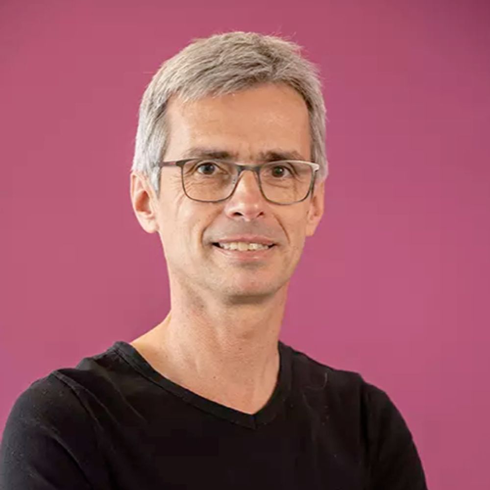
Pierre-François Lenne
@pflenne.bsky.social
920 followers
220 following
43 posts
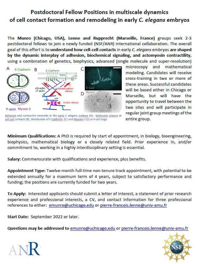
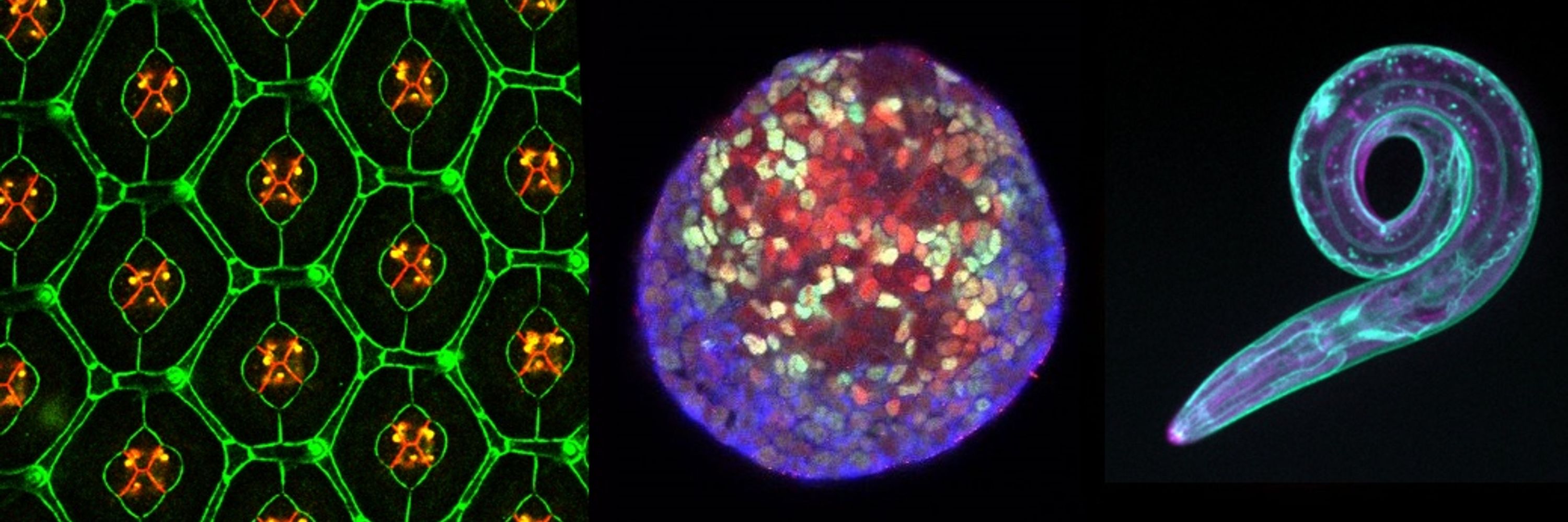
Biophysicist interested in cell dynamics and tissue morphogenesis
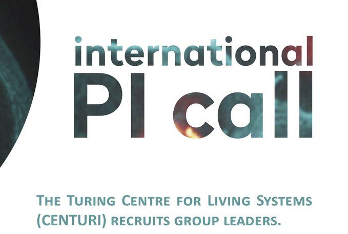
at IBDM and Turing Center for Living Systems
Group: https://www.morphotiss.org/
https://www.ibdm.univ-mrs.fr/physical-approaches-to-cell-dynamics/
https://centuri-livingsystems.org
Posts
Media
Videos
Starter Packs
Pinned
Reposted by Pierre-François Lenne

Reposted by Pierre-François Lenne