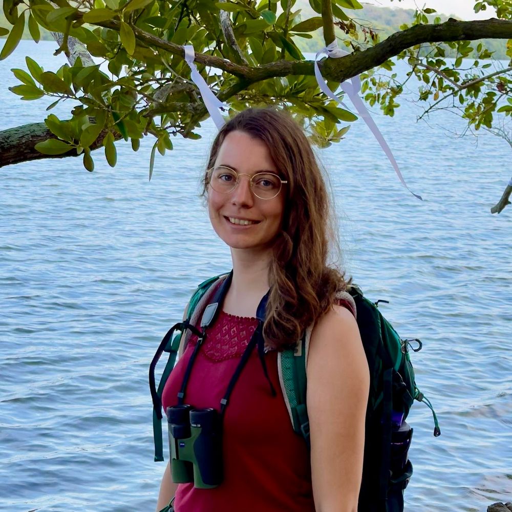
Susanne Babl
@susannebabl.bsky.social
85 followers 100 following 9 posts
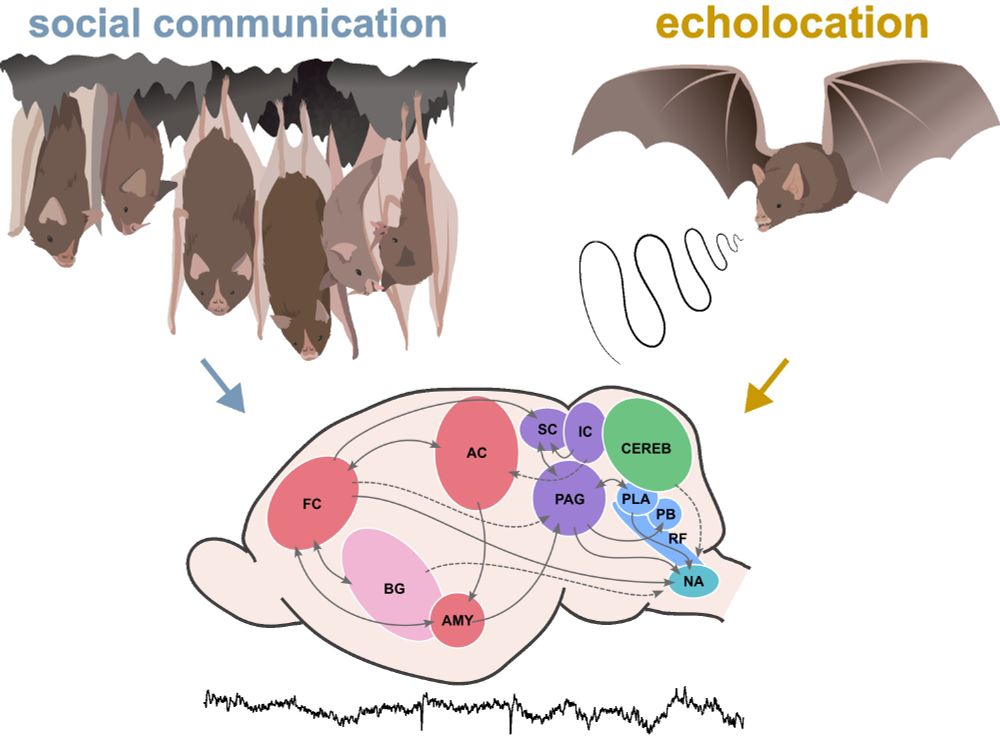
Neuroethologist - bats and brains 🦇🦇🧠 Postdoc at FU Berlin
Posts Media Videos Starter Packs
susannebabl.bsky.social
Last week, I moved from Frankfurt to Berlin and started my new postdoc position at Freie Uni with @talking-bat.bsky.social and @ycabral.bsky.social 🥳
I am super excited to set up our new lab for more work on bats and their brain+behavior! 🦇🧠🧠
Reposted by Susanne Babl
berlinbatlab.bsky.social
New paper out!
We found that in pipistrelle bats, social vocalizations carry a stronger phylogenetic signal than echolocation calls, suggesting the former evolve more slowly, while the latter remains flexible and shaped by the environment.
www.sciencedirect.com/science/arti...
susannebabl.bsky.social
Team effort by Ava Kiai, @talking-bat.bsky.social , Francisco García-Rosales and me :)
susannebabl.bsky.social
Are you interested in bat vocalizations and the neural circuits that give rise to their emission?🦇🎶🧠
We reviewed the recent literature on neural activity underlying echolocation and communication calls across multiple bat species in Annals of the New York Academy of Sciences:
doi.org/10.1111/nyas...
Reposted by Susanne Babl
ahanaaurora.bsky.social
Ever wondered how baby bats learn to sing? 🦇🎶
Turns out, their moms support their practice! Our study shows that maternal feedback leads to:
More vocal practice
Higher song syllable versatility
More mature song syllables
🔗https://elifesciences.org/articles/99474
elifesciences.org/digests/9947...
Mother-pup pair of the Greater sac-winged bat in their day roost.
Reposted by Susanne Babl
plosbiology.org
#Hippocampus & PFC interact to support spatial #WorkingMemory, but are dHPC & vHPC functionally redundant in this? @susannebabl.bsky.social @torfisigurdsson.bsky.social show that both regions contribute differentially to spatial WM & coding of spatial info by PFC @plosbiology.org 🧪 plos.io/4cXyddO
Top left: Schematic of virus injection and optic fiber implantation in dHPC (left) or vHPC (right) with electrodes implanted in the PFC to record neuronal activity during hippocampal silencing. Right: Example coronal sections showing ArchT-GFP expression in dHPC (top) and vHPC (bottom) with optic fiber placements outlined by dashed white lines. Bottom left: Task schematic. In the sample phase (left), mice were guided to enter one of the two goal arms to receive a reward. After a delay phase in the start box (middle), mice could choose between both goal arms but only received a reward in the goal arm opposite to the one visited in sample (right).
susannebabl.bsky.social
Our results indicate distinct contributions of the two hippocampal poles in spatial working memory and in communicating relevant spatial information to the prefrontal cortex.
susannebabl.bsky.social
Interestingly, prefrontal representations of the animal’s goal location during encoding was disrupted only when the ventral HPC was inactivated. Inactivation of the dorsal HPC had no effect on these spatial goal codes.
susannebabl.bsky.social
Then, we looked at prefrontal activity when either dorsal or ventral HPC were inactivated. We found that while neurons in the prefrontal cortex still represented the animal’s position in the maze, spatial codes were altered compared to trials with a functioning HPC.
susannebabl.bsky.social
However, the ventral HPC is only necessary in the encoding phase. Neither ventral nor dorsal HPC were needed to maintain the location in memory for a few seconds.
susannebabl.bsky.social
First, we found that the dorsal and the ventral HPC play distinct roles in supporting spatial working memory: the dorsal HPC is necessary during two critical phases of the task, namely during encoding a spatial location and while retrieving this information.
Reposted by Susanne Babl