Teun Huijben
@teunhuijben.bsky.social
1.8K followers 490 following 55 posts
Scientist/PostDoc with Loïc Royer at Chan Zuckerberg Biohub, San Francisco 🔬 image analysis | localization microscopy | machine learning, 🇳🇱 by bike 🚲 📍San Francisco
Posts Media Videos Starter Packs
Pinned
teunhuijben.bsky.social
#HiSciSky, time for an introduction! I work with Loïc Royer at the CZ Biohub in San Francisco. My work focuses on image analysis, light-sheet microscopy, and AI. I did my PhD in SMLM, so expect a mix of localization microscopy, cell tracking, light-sheet movies, data viz, and super-resolution!💡🧪🔬
teunhuijben.bsky.social
Had a great time in Janelia this summer developing #geff with this amazing group of cell tracking enthousiasts and software engineers! #geff is a file format for tracking data allowing universal cross-tool communication for a wealth of tracking tools, amongst others our #ultrack and #inTRACKtive 🚀
msschwartz21.bsky.social
#geff v1 is released! 🎉 GEFF is a zarr-based Graph Exchange File Format with special support for tracking applications in Python and Java. Thank you to all the amazing developers who contributed at the Janelia 2025 Trackathon! liveimagetrackingtools.org/geff/
teunhuijben.bsky.social
🍾What a day 🥳 Two papers of the Royer lab in Nature Methods today, including #inTRACKtive! Check out Loïc's thread below for the details ⬇️

Over the next days, I will share the exciting new features we have added since the original preprint. Stay tuned!
loicaroyer.bsky.social
🧬🔬🧪 🎉 Our team at @czbiohub is thrilled to share TWO companion papers out today in @NatureMethods!
📦 Ultrack — robust, scalable nD cell tracking
🌐 inTRACKtive — a beautiful, open-source web viewer for lineage exploration
Let’s dive in! 👇
www.nature.com/articles/s41...
Reposted by Teun Huijben
jytinevez.bsky.social
Saying goodbye to beautiful @hhmijanelia.bsky.social, after an intense two weeks trackhathon. Organized by legends Caroline Malin-Mayor and Morgan Schwartz in the @janfunkey.bsky.social.

It has been awesome and productive. Thank you!!
teunhuijben.bsky.social
Enjoying two weeks of cell tracking tools, file formats, and collaboration, all at the beautiful Janelia Research Campus. What a place! 🤩🔬🧪 @hhmijanelia.bsky.social
Janelia research campus - Virginia
Reposted by Teun Huijben
aafkegros.bsky.social
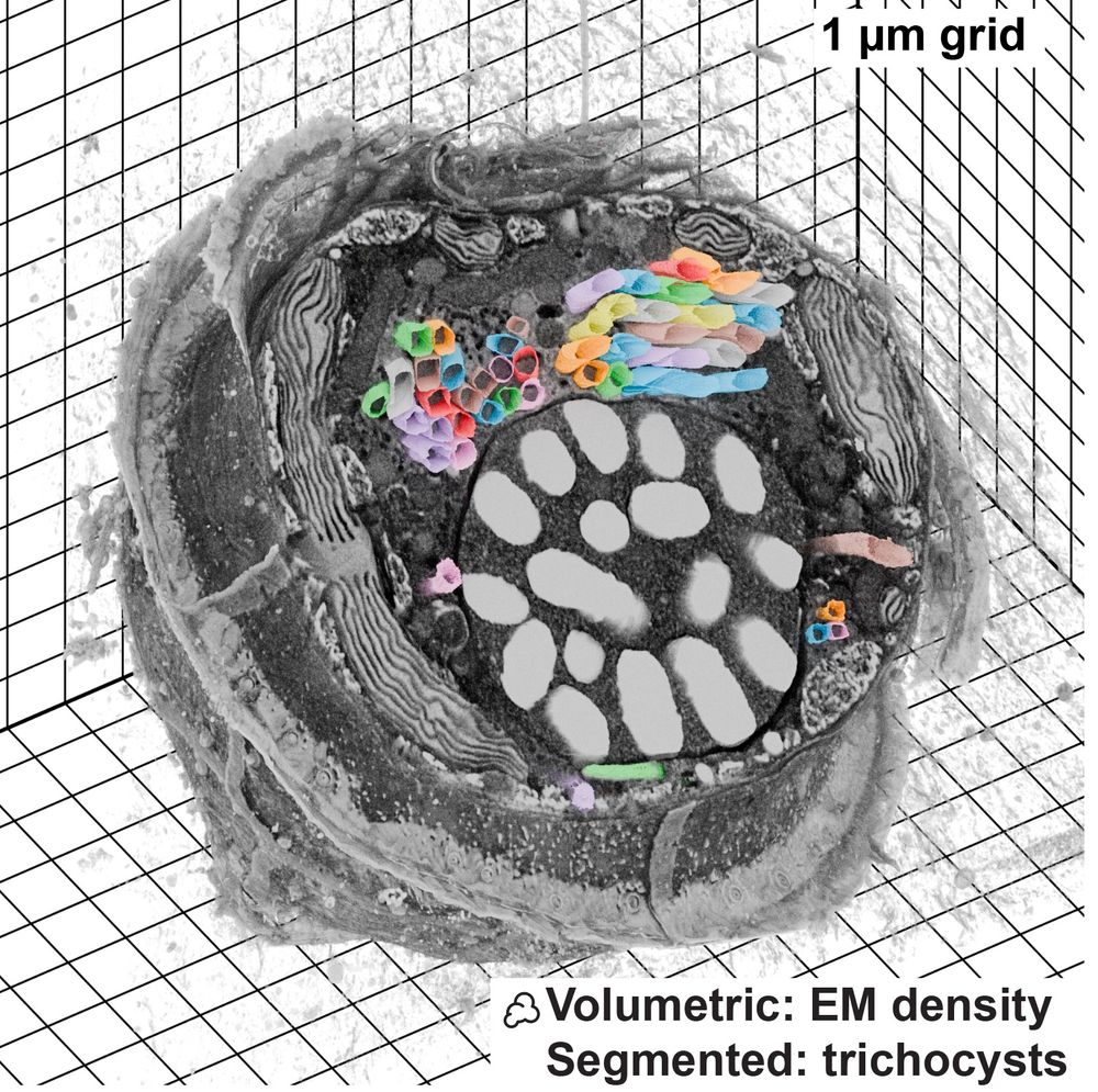
Microscopy Nodes is now up on bioRxiv! 🚀

This is a Blender extension that seamlessly integrates and visualizes 3D microscopy data (TIF & @zarr.dev).

High-quality volume rendering for anyone, in both EM and fluorescence, regardless of computational expertise! 🔬

www.biorxiv.org/content/10.1...
FIB-SEM dataset visualized with Microscopy Nodes, data from Mocaer et al 2023
teunhuijben.bsky.social
Little Nanobiology reunion in San Diego. Great to see multiple professors/alumni at the Cell Bio conference! 🙌

@tudelft.bsky.social #nanobiology #ASCB24 #CellBio2024
liedewijlaan.bsky.social
Nanobiology Rocks! #ASCB2024
Poster ASCB two nanobiologists
teunhuijben.bsky.social
Happy to be in sunny☀️San Diego for ASCB Cell Bio! Tomorrow I will present #Ultrack and #inTRACKtive (poster B513) at 11:15 am. Swing by the [email protected]
booth (#119) for live demos of these tools + #napari & #zebrahub 🐠 Looking forward to connect!🤩🧪🔬

#CellBio2024 #ASCB24 #celltracking
Sunny San Diego view
teunhuijben.bsky.social
Join us next Friday at UC San Diego for a day full of microscopy 💡🔬, mass spec, and AI! Shout out to @manorlaboratory.bsky.social for putting this together! I will present our recent work on cell tracking algorithms and visualization with #Ultrack and #inTRACKtive🧪

#CellBio2024
manorlaboratory.bsky.social
Re-upping this - please register if you're in San Diego this Friday and want to hear about the latest advances in cryoEM/cryoET, mass spec, live imaging, and AI!
tinyurl.com/ASCBSANDBOX
teunhuijben.bsky.social
Hi Eric, could you add me to the Science 🧪 feed? I am employed at the Chan Zuckerberg Biohub in San Francisco working at the interface of developmental biology, microscopy, and machine learning.

Google Scholar: scholar.google.com/citations?us...

Orcid: orcid.org/0000-0002-89...
teunhuijben.bsky.social
Congrats on the paper! Also, it’s time to bring Sjoerd Stallinga over to the BlueSky-verse ;)
Reposted by Teun Huijben
heilemannlab.bsky.social
We modified our STED🔬to make it more efficient ('smart') to acquire high-resolution data of small and rare objects in a 3D cell volume. This enabled collecting a large amount of data in a fully automated fashion. With that, we visualized the ulra-structure a small nuclear body, the paraspeckle👇🏻(1/3)
teunhuijben.bsky.social
10/ Thanks to all co-authors: Sarojini Mahajan, Masih Fahim, Peter Zijlstra, Rodolphe Marie and Kim I. Mortensen🙌. And of course our #Horizon2020 #MSCActions SuperCol ITN network! 🥳
Point-spread function deformations unlock 3D localization microscopy on spherical nanoparticles
teunhuijben.bsky.social
9/ In addition to the metal nanoparticles (gold 🟠), Masih Fahim’s work showed that the same holds for dielectric (polystyrene ⚪️) particles, allowing us to reconstruct the distribution of single DNA molecules on the nanoparticle surface 🧬
 In addition to the metal nanoparticles (gold 🟠), Masih Fahim’s work showed that the same holds for dielectric (polystyrene ⚪️) particles, allowing us to reconstruct the distribution of single DNA molecules on the nanoparticle surface 🧬
teunhuijben.bsky.social
8/ In this way, we can map out the full surface. We show that the surface functionalization is patchy and heterogeneous between particles. This will allow studying the surface functionalization at the single-particle level to design and produce the nanoparticles of the future.
teunhuijben.bsky.social
7/ Fitting the experimental images with our new analytical PSF model, we obtain the coordinates of the emitters relative to the nanoparticle surface. Allowing us to map the position of the DNA strands that engaged in DNA-PAINT, with <5nm precision!
Fitting the experimental images with our new analytical PSF model, we obtain the coordinates of the emitters relative to the nanoparticle surface. Allowing us to map the position of the DNA strands that engaged in DNA-PAINT, with <5nm precision!
teunhuijben.bsky.social
6/ To showcase the fitting approach, we perform DNA-PAINT on DNA-coated 100nm gold nanoparticles, resulting in a zoo of exotic PSFs!
Zoo of exotic PSF shapes resulting from DNA-PAINT on gold spherical nanoparticles
teunhuijben.bsky.social
5/ We developed the first-ever analytical PSF model for a fluorophore near a spherical nanoparticle, of any size, material, and composition. Our model is 4 orders of magnitude faster than numerical calculations, allowing us to use it directly to fit experimental data.
We developed the first-ever analytical PSF model for a fluorophore near a spherical nanoparticle, of any size, material, and composition. Our model is 4 orders of magnitude faster than numerical calculations, allowing us to use it directly to fit experimental data.
teunhuijben.bsky.social
4/ The PSF doesn’t always deform in the same way, but the PSF shape depends on the fluorophore’s position relative to the nanoparticle. This means that the PSF shape encodes information about the fluorophore’s position, which we can use to our advantage during localization.
teunhuijben.bsky.social
3/ A single (freely rotating) fluorophore creates a symmetric PSF, which we conventionally fit with a 2D Gaussian. However, the presence of a nanoparticle deforms the PSF, resulting in significant biases when simply fitted with a Gaussian.
A single (freely rotating) fluorophore creates a symmetric PSF, which we conventionally fit with a 2D Gaussian. However, the presence of a nanoparticle deforms the PSF, resulting in significant biases when simply fitted with a Gaussian.
teunhuijben.bsky.social
2/ Nanoparticles are important for biosensing, drug delivery, and cancer therapy. The functionality of the nanoparticle crucially depends on the distribution and number of functional groups on their surface, which can ideally be studied with localization microscopy🔬
Nanoparticles are important for biosensing, drug delivery, and cancer therapy.
teunhuijben.bsky.social
PSFs are just Gaussians, right??

Not when your fluorophore💡is close to a nanoparticle 🟠!
Excited to share our recent ACS Nano paper 🥳 in which we present a novel PSF-fitting approach for localization microscopy near nanoparticles.

pubs.acs.org/doi/full/10....

Main findings below in🧵[1/10]
teunhuijben.bsky.social
8/ In this way, we can map out the full surface. We show that the surface functionalization is patchy and heterogeneous between particles. This will allow studying the surface functionalization at the single-particle level to design and produce the nanoparticles of the future.
We show that the surface functionalization is patchy and heterogeneous between particles.
teunhuijben.bsky.social
7/ Fitting the experimental images with our new analytical PSF model, we obtain the coordinates of the emitters relative to the nanoparticle surface. Allowing us to map the position of the DNA strands that engaged in DNA-PAINT, with <5nm precision!
Fitting the experimental images with our new analytical PSF model, we obtain the coordinates of the emitters relative to the nanoparticle surface.
teunhuijben.bsky.social
6/ To showcase the fitting approach, we perform DNA-PAINT on DNA-coated 100nm gold nanoparticles, resulting in a zoo of exotic PSFs!
DNA-PAINT on DNA-coated 100nm gold nanoparticles results in a zoo of exotic PSFs
teunhuijben.bsky.social
5/ We developed the first-ever analytical PSF model for a fluorophore near a spherical nanoparticle, of any size, material, and composition. Our model is 4 orders of magnitude faster than numerical calculations, allowing us to use it directly to fit experimental data.
Schematics of the analytical and numerical approaches for calculating the PSF for an emitter (red dot) in close proximity to an NP (gold sphere) in water on top of a glass coverslip (blue rectangle). In the analytical model, we directly and analytically derive the electromagnetic fields everywhere in the system. In the numerical approach, the fields are computed in a finite element manner on the discretized system