Centre for the Cellular Microenvironment
@theglasgowcemi.bsky.social
1.7K followers 2K following 92 posts
We are a Glasgow-based multidisciplinary team of bio- and cell engineers. We focus on understanding the interactions between materials, proteins and cells to gain insight into engineering cell behaviour and translating technologies into better healthcare.
Posts Media Videos Starter Packs
Reposted by Centre for the Cellular Microenvironment

carstenschulte4.bsky.social
The core team of the Tenovus Scotland-funded Small Pilot Grant project "Leveraging mechanobiology for the optimisation of in vitro muscle-related disease models" is on the case😊, with a strong 4th&5th year students-driven component! @unistrathclyde.bsky.social @theglasgowcemi.bsky.social
Reposted by Centre for the Cellular Microenvironment
prism-livingtissues.eu
Alexandre, from @theglasgowcemi.bsky.social, will share how our team is developing “symbiotic engineered living materials” — 3D constructs where genetically engineered helper cells detect metabolic cues from mesenchymal stem cells and respond by secreting lineage-specific growth factors.
Reposted by Centre for the Cellular Microenvironment
carstenschulte4.bsky.social
We are very happy 😄 that our Rheinland-Pfalz & Scotland exchange project started, jointly awarded by @sulsa.bsky.social and JGU Scotland Hub, sponsored by the governments of 🏴󠁧󠁢󠁳󠁣󠁴󠁿 & Rheinland-Pfalz. 1/2
@unistrathclyde.bsky.social @theglasgowcemi.bsky.social @imbmainz.bsky.social @unimainz.bsky.social
Reposted by Centre for the Cellular Microenvironment
uofglasgow.bsky.social
As we mark #PrideMonth, we take this moment to celebrate LGBTQ+ identities and also reflect on the ongoing work to make our universities places where every member can thrive.

Thank you to colleagues for co-authoring this powerful piece on Rainbow Office Hours ➡️ gla.ac/3Zio6uv

#OneTeamUofG 🏳️‍🌈 🏳️‍⚧️
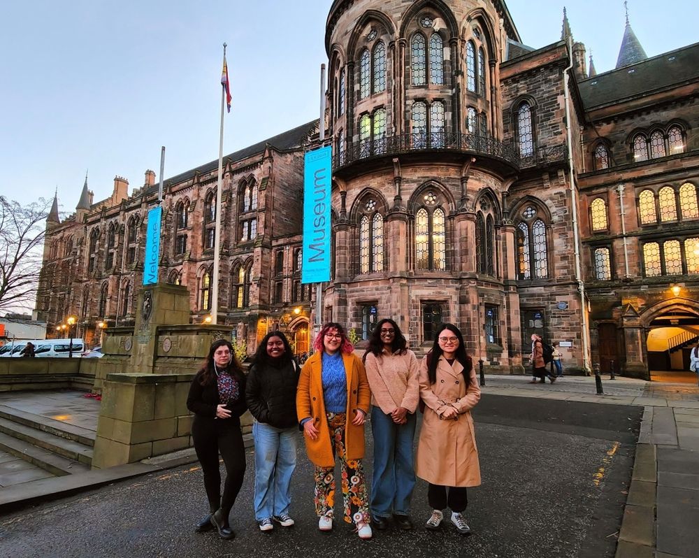
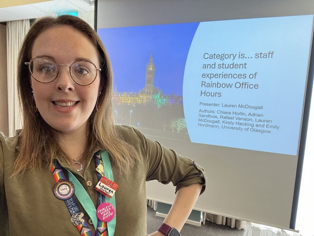
University of Glasgow illuminated in rainbow pride flag colours Queer teachers are living proof of queer futures - created by Adrian A group of people standing outside of the univeristy of glasgow main building Lauren stands in front of a presentation screen, it's titled: Category is... staff and student experiences of rainbow office hours.
Reposted by Centre for the Cellular Microenvironment
giudica.bsky.social
New review paper out! Proud to co-author with @optophysakhil.bsky.social and @gailmcconnell.bsky.social a deep dive into Fourier Ptychographic Microscopy (FPM), from physics to cutting-edge algorithms & medical imaging applications. onlinelibrary.wiley.com/doi/epdf/10....
Fourier ptychography microscopy for digital pathology
onlinelibrary.wiley.com
Reposted by Centre for the Cellular Microenvironment

carstenschulte4.bsky.social
A little more than a year that @carmeloferrai.bsky.social and I published our review "Mechanotransduction in stem cells"🤯! I'm happy that it was received well 😃! Stay tuned, more to come in this direction in a not too far future😎.
carmeloferrai.bsky.social
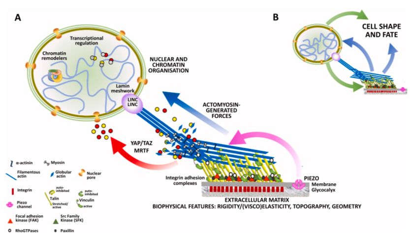
EpiSci - Hi there! Our review “Mechanotransduction in stem cells” is finally published. With @carstenschulte4.bsky.social we provided a comprehensive picture, that aims to cover the whole mechanotransductive pathway from the cell microenvironment to the nucleus. 1/3 pubmed.ncbi.nlm.nih.gov/38729084/
Reposted by Centre for the Cellular Microenvironment
giudica.bsky.social
At the Center of the Cellular Microenvironment’s impact workshop. Really enjoying the space to think together about where cell engineering and mechanobiology are headed. @theglasgowcemi.bsky.social @unistrathclyde.bsky.social
theglasgowcemi.bsky.social
A chat with Prof. Viola Vogel (Department of Health Sciences and Technology, ETH Zürich), whose pioneering work in mechanobiology has been key to show that proteins act as mechano-chemical switches which can be exploited in the treatment of various diseases.

podcasts.apple.com/gb/podcast/m...
Making progress in mechanobiology (with Prof Viola Vogel)
Podcast Episode · The CeMi Podcast · 20/03/2025 · 23m
podcasts.apple.com
theglasgowcemi.bsky.social
Congratulations to Anna Maria Kapetanaki who passed her PhD viva this week! 🎉 🥳 💓:
Reposted by Centre for the Cellular Microenvironment
carstenschulte4.bsky.social
Thanks, I'm happy that you like it😊.
Indeed, another critical component is molecular clutch force loading:

bsky.app/profile/cars...
carstenschulte4.bsky.social
I delved even deeper into this a few years later, further highlighting the importance of talin-mediated molecular clutch force loading in this process:

👉 www.sciencedirect.com/science/arti...
Reposted by Centre for the Cellular Microenvironment
carstenschulte4.bsky.social
...can involve also other cell adhesion receptors (e.g., GPI-anchored, such as uPAR, or maybe syndecans, DDRs?) 2/2:
Reposted by Centre for the Cellular Microenvironment
carstenschulte4.bsky.social
Consistently, some years later (2019), Changede et al. (Sheetz group) showed the existence of integrin nanoclusters bridges build up by activated, but unliganded, integrins. The binding to the thin matrix fibres was mediated by integrins in this case, but it's intriguing to think that this 1/2
Reposted by Centre for the Cellular Microenvironment
carstenschulte4.bsky.social
I delved even deeper into this a few years later, further highlighting the importance of talin-mediated molecular clutch force loading in this process:

👉 www.sciencedirect.com/science/arti...
Reposted by Centre for the Cellular Microenvironment
carstenschulte4.bsky.social
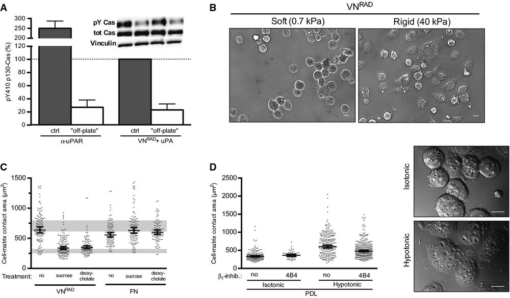
This ligand binding-independent integrin signalling was driven by membrane tension. Indeed, the induction of this non-canonical integrin signalling was not restricted to uPAR/vitronectin interaction.
This work introduced the cell membrane as a critical component of the molecular clutch.
Figure 7. uPAR/VN signalling requires cell adhesion, ECM rigidity and membrane tension
A.
uPAR‐induced signalling requires cell attachment. Cells expressing uPART54A were seeded on an anti‐uPAR monoclonal antibody (α‐uPAR) or on VNRAD (with uPA) for 30 min. For ‘off‐plate’ experiments, cells were incubated in suspension with these components. Phosphorylation of p130Cas SD is shown in percentage of the positive control, that is the same cells seeded on VNRAD in the presence of uPA (n ≥ 3, mean ± SEM). Representative blots are shown.
B.
uPAR‐induced cell spreading requires adhesion to a rigid matrix. Representative images of 293 cells expressing uPART54A plated in presence of uPA on hydrogels coated with VNRAD at different rigidity (soft: 0.7 kPa, Rigid: 40 kPa) for 30 min. Scale bar: 10 μm.
C.
Effect of plasma membrane relaxation on uPAR or integrin‐induced cell spreading. 293 cells expressing uPART54A were seeded on FN or VNRAD (with uPA) in presence of sucrose (150 mM) or deoxycholate (0.4 mM) for 30 min. Cell‐matrix contact area was quantified. Measurements represent at least 50 cells with indicated geometrical means ± 95% CI, in at least two independent experiments. The grey bars represent the range of cell area of not spread or fully spread uPART54A cells based on 95% CI on poly‐D‐lysine or on VN with uPA, respectively (Fig 2B).
D.
Increased membrane tension induces integrin‐dependent cell spreading. Cells were seeded on PDL for 30 min in presence or absence of 4B4. Where indicated, an equal volume of hypotonic buffer (H2O with 1 mM MgCl2 and 1.2 mM CaCl2) or isotonic (medium) buffer was added to increase plasma membrane tension. Cell‐matrix contact area was assayed and quantified. Measurements represent at least 50 cells with indicated geometrical means ± 95% CI, in at least two independent experiments. Representative DIC images of cells on PDL treated with isotonic or hypotonic medium are shown. Scale bar: 10 μm.
Reposted by Centre for the Cellular Microenvironment
carstenschulte4.bsky.social
This study assessed the possibility of ligand-independent adhesion signalling by integrins, which was a quite controversial hypothesis at the time (maybe still is).
We found that e.g. uPAR-mediated adhesion to vitronectin can induce integrin signalling that does not require integrin ligand binding:
theglasgowcemi.bsky.social
Interesting 🧵on a classic mechanotransduction paper of one of our members introducing the concept of ligand binding-independent integrin signalling driven by membrane tension:
theglasgowcemi.bsky.social
The abstract submission deadline of BioMedEng2025 @biomedgen25.bsky.social has been extended. You still have the chance to submit until the 19th of May:

👉 biomedeng.org/biomedeng25/
Reposted by Centre for the Cellular Microenvironment
marcocantini.bsky.social
In this work my team and co-workers demonstrated a purely mechanically regulated route for #StemCell #Chondrogenesis via engineering of the hydrogels #viscous nature. No #growthfactors needed. Conducted @uofglasgow.bsky.social @theglasgowcemi.bsky.social Funded @ukri.org
Reposted by Centre for the Cellular Microenvironment
marcocantini.bsky.social
Great to see that our work on #viscoelastic #hydrogels for #cartilage #TissueEngineering was one of the most-read papers in Advanced Healthcare Materials @mattjwalker.bsky.social @delphe.bsky.social @manlio-tassieri.bsky.social
Check it out here doi.org/10.1002/adhm... 🧪
Top Viewed Article in Advanced Healthcare Materials in 2023
Reposted by Centre for the Cellular Microenvironment
carstenschulte4.bsky.social
Maybe the opportunity of your life, become the Head of the wonderful Department of Biomedical Engineering @unistrathclyde.bsky.social in Glasgow 🏴󠁧󠁢󠁳󠁣󠁴󠁿 :
👉 www.nature.com/naturecareer...
Reposted by Centre for the Cellular Microenvironment