Matt Tyska
@tyskalabactual.bsky.social
1.3K followers 1.2K following 24 posts
Interested in how the cytoskeleton controls cell morphology and function; yes to microscopy. 
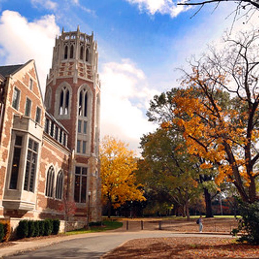
https://lab.vanderbilt.edu/tyska-lab/
Posts Media Videos Starter Packs
Reposted by Matt Tyska
stefvass.bsky.social
💥 Quick thread on some stunning actin filament swirls🌪️ spotted under platinum replica EM, often forming nest-like structures around caveolae🏐
#ActinAesthetics #ElectronMicroscopy
Reposted by Matt Tyska
vandybiomed.bsky.social
Happy Friday! Here to bring a bit of brightness to your day are a few images by Julissa Burgos, one of our graduate students in the Tyska lab here at Vanderbilt. Gorgeous!! 🔬
@yuyi106.bsky.social
@tyskalabactual.bsky.social
#fluorescencefriday #cellbiology
tyskalabactual.bsky.social
Also Monday at #cellbio2024! Julissa (@yuyi106.bsky.social) will present her studies on the role of an I-BAR protein (IRTKS) in the pathogenic mechanism of Enterohemorrhagic E. coli 🦠🧫🔬 P2352. B677. @vucellimaging.bsky.social
Reposted by Matt Tyska
binyammogessie.bsky.social
This is couple of years old since I rarely have time to sit at a scope these days but I was inspired by the end of year #FluorescenceFriday posts on hear and thought I should drop on y’all my Eye of Sauron, aka a mouse oocyte containing nuclear actin filaments #actin #meiosis #oocytes
Reposted by Matt Tyska
zlehmann.bsky.social
Happy #FluorescenceFriday! These are polarized epithelial cells with microvilli on their apical surface! Captured using the #Nikon NSPARC, and color-coded by depth #actin #microscopy @tyskalabactual.bsky.social
Reposted by Matt Tyska
francescoferraro.bsky.social
This one is for the holiday season
tyskalabactual.bsky.social
yes and it’s such a tiny little thing!
tyskalabactual.bsky.social
Thought Spef1 one was some ciliary protein? Check out Dr. Rocio‘s (@rtpastrana.bsky.social) poster on how Spef1 shapes epithelial monolayers, no cilia needed 🤩. Tuesday at #cellbio2024, P2439, B46. @vucellimaging.bsky.social
Reposted by Matt Tyska
operkins.bsky.social
Have you always wondered how microvilli form? Where DOES that actin come from to form the core bundle? Come to the #BuildingTheCell (Part 1) subgroup tomorrow morning at 8:45 to find out! Room 29C. #CellBio2025

@tyskalabactual.bsky.social
tyskalabactual.bsky.social
Also, also on Monday at #cellbio2024 I get to present Olivia’s (@operkins.bsky.social) unexpected discoveries on the link between clathrin-mediated endocytosis and the growth of epithelial microvilli 🤯
P1831. B137.
@vucellimaging.bsky.social
tyskalabactual.bsky.social
Also Monday at #cellbio2024! Julissa (@yuyi106.bsky.social) will present her studies on the role of an I-BAR protein (IRTKS) in the pathogenic mechanism of Enterohemorrhagic E. coli 🦠🧫🔬 P2352. B677. @vucellimaging.bsky.social
tyskalabactual.bsky.social
Posters on tuft cells are about as rare as tuft cells, but at #cellbio2024 on Monday, @jen-silverman.bsky.social presents “P1718. Organization of a cytoskeletal superstructure in the apical domain of intestinal tuft cells. B23” Yes! those are gigantic actin bundles 🤯
@vucellimaging.bsky.social
tyskalabactual.bsky.social
Synchronized filopodial formation for
#FluorescenceFriday 💥
#CellBiology #Microscopy @vucellimaging.bsky.social
tyskalabactual.bsky.social
Intestinal #organoid goes rogue and flips polarity 🤯
@vucellimaging.bsky.social
#epithelial #cellbiology #microscopy
Reposted by Matt Tyska
vucellimaging.bsky.social
It's that time of year. Confocal + Christmas = Xmas Tree, Ymas Tree, and our favorite Zmas tree. #microscopy #confocal #imaging
Reposted by Matt Tyska
pjsaez.bsky.social
As promised, here a fast acquisition of a migrating cell videoed under a microscopy, look those membrane dynamics @ the leading edge !

Bonus: nuclear deformation once the cell rear snaps.
(Fluo nucleus, for the #fluorescencefriday)

@focalplane.bsky.social @cellcommlab.bsky.social #CellMigration 🧪🔬
Reposted by Matt Tyska
microtubule.bsky.social
Highlighting our recent paper on how we think doublecortin (green) contributes to growth cone biomechanics in developing neurons for #FluorescenceFriday

www.cell.com/current-biol...
Reposted by Matt Tyska
lrcap.bsky.social
When you’re imaging and your sample takes a Pikachu!
Having fun playing around with our new #Nikon NSPARC for #FluorescenceFriday
#microscopy #myosin #epithelial #cellbiology #meme
@tyskalabactual.bsky.social
tyskalabactual.bsky.social
O is for #organoid

@vucellimaging.bsky.social
#microscopy #epithelial #cellbiology
tyskalabactual.bsky.social
The real IFT…
intra💥filopodial💥transport.
@vucellimaging.bsky.social
#cytoskeleton #biology #microscopy
Reposted by Matt Tyska
axonsalex.bsky.social
Starting the week strong! Cheering myself up for the next challenges with 15 min of axonal growth cone dynamic madness. F-actin (cyan) and membrane (magenta) show their dance. One image taken every 10 s.🧠🔬
#MicroscopyMonday #LiveImaging #Neuroscience #Microscopy