Blake Ushijima, Ph.D.
@vibriosoup.bsky.social
960 followers
1.3K following
130 posts
Assistant Prof at UNCW | Microbiology | Vibrio coralliilyticus enthusiasts | Coral pathogens & probiotics 🪸🦠 | #SCTLD | Comments are my own | Hawaii born | he/him
https://www.ushijima-lab.com/
Posts
Media
Videos
Starter Packs

Reposted by Blake Ushijima, Ph.D.


Reposted by Blake Ushijima, Ph.D.
Reposted by Blake Ushijima, Ph.D.


Reposted by Blake Ushijima, Ph.D.


Reposted by Blake Ushijima, Ph.D.


Reposted by Blake Ushijima, Ph.D.


Reposted by Blake Ushijima, Ph.D.

Reposted by Blake Ushijima, Ph.D.


Reposted by Blake Ushijima, Ph.D.
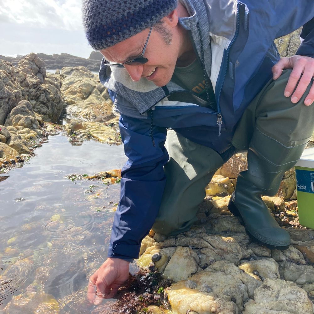
Matt Bracken
@brackenlab.bsky.social
· Jul 6

Reposted by Blake Ushijima, Ph.D.