


Volume Imaging Australia
@volumeimagingaus.bsky.social
Special Interest Group of Australian Microscopy & Microanalysis Society (AMMS)
Pinned

Sound the trumpets, the 2025 Light Microscopy Australia & Volume Imaging Australia Imaging Awards are now open. Huzzah! Submit your spiffing light microscopy, volume electron microscopy and x-ray microscopy images for your chance to win some cold hard cash! Details via the QR code below 😀🔬
Sound the trumpets, the 2025 Light Microscopy Australia & Volume Imaging Australia Imaging Awards are now open. Huzzah! Submit your spiffing light microscopy, volume electron microscopy and x-ray microscopy images for your chance to win some cold hard cash! Details via the QR code below 😀🔬
October 20, 2025 at 6:11 AM
Sound the trumpets, the 2025 Light Microscopy Australia & Volume Imaging Australia Imaging Awards are now open. Huzzah! Submit your spiffing light microscopy, volume electron microscopy and x-ray microscopy images for your chance to win some cold hard cash! Details via the QR code below 😀🔬
Reposted by Volume Imaging Australia

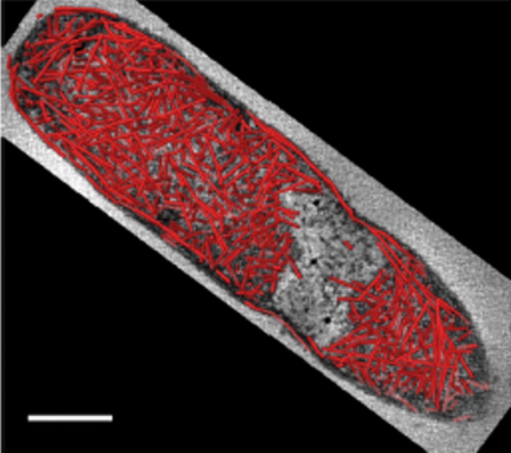
Volume EM meets synthetic biology
Matthew Lee, Judith Mantell, Martin Warren, Paul Verkade and colleagues use serial section Transmission Electron Tomography (ss-ET) to visualise synthetic ‘cytoscaffolds’ in bacteria.
focalplane.biologists.com/2025/06/27/v...
@volumeem1.bsky.social 🔬
Matthew Lee, Judith Mantell, Martin Warren, Paul Verkade and colleagues use serial section Transmission Electron Tomography (ss-ET) to visualise synthetic ‘cytoscaffolds’ in bacteria.
focalplane.biologists.com/2025/06/27/v...
@volumeem1.bsky.social 🔬
volume EM visualisation of synthetic scaffolds in bacteria - FocalPlane
volume EM visualisation of synthetic scaffolds in bacteria - Volume EM
focalplane.biologists.com
July 10, 2025 at 3:16 PM
Volume EM meets synthetic biology
Matthew Lee, Judith Mantell, Martin Warren, Paul Verkade and colleagues use serial section Transmission Electron Tomography (ss-ET) to visualise synthetic ‘cytoscaffolds’ in bacteria.
focalplane.biologists.com/2025/06/27/v...
@volumeem1.bsky.social 🔬
Matthew Lee, Judith Mantell, Martin Warren, Paul Verkade and colleagues use serial section Transmission Electron Tomography (ss-ET) to visualise synthetic ‘cytoscaffolds’ in bacteria.
focalplane.biologists.com/2025/06/27/v...
@volumeem1.bsky.social 🔬
If, like us, segmentation of microscopy images is your jam or if you want to learn more about how segmentation could be your jam, then our upcoming webinar is for you!
Segment Anything for Microscopy - Constantin Pape
8 July, 4:00 – 5:00 PM AEST
Registration and details: tinyurl.com/wt7c4avu
Segment Anything for Microscopy - Constantin Pape
8 July, 4:00 – 5:00 PM AEST
Registration and details: tinyurl.com/wt7c4avu

VIA Webinar: Segment Anything for Microscopy - Microscopy Australia
Join us for this Volume Imaging Australia webinar as we explore ‘Segment Anything for Microscopy’ (μSAM), a versatile tool for segmentation with Prof. Constantin Pape. Abstract Accurate segmentation o...
micro.org.au
June 27, 2025 at 4:42 AM
If, like us, segmentation of microscopy images is your jam or if you want to learn more about how segmentation could be your jam, then our upcoming webinar is for you!
Segment Anything for Microscopy - Constantin Pape
8 July, 4:00 – 5:00 PM AEST
Registration and details: tinyurl.com/wt7c4avu
Segment Anything for Microscopy - Constantin Pape
8 July, 4:00 – 5:00 PM AEST
Registration and details: tinyurl.com/wt7c4avu
An excellent and eye-opening new case study on the awesomeness of SBF-SEM!

Discover how SBF-SEM (Serial Block-Face Scanning Electron Microscopy), a vEM technique, is being used to unveil new perspectives on corneal development.
Check out our new case study: tinyurl.com/54pxp6ku
Authors: Rob Young, Kiranjit Bains, Phil Lewis and Andrew Quantock - Cardiff University, UK
Check out our new case study: tinyurl.com/54pxp6ku
Authors: Rob Young, Kiranjit Bains, Phil Lewis and Andrew Quantock - Cardiff University, UK

New insights into corneal development from vEM - FocalPlane
New insights into corneal development from vEM - Volume EM
tinyurl.com
June 27, 2025 at 4:21 AM
An excellent and eye-opening new case study on the awesomeness of SBF-SEM!
A new version of Empanada, the fantastic deep learning mitochondria segmentation tool, has just been released!
It also includes expanded tools for other organelles: DropNet for Lipid Droplets and NucleoNet for Nuclei. Learn more here: empanada.readthedocs.io/en/latest/
It also includes expanded tools for other organelles: DropNet for Lipid Droplets and NucleoNet for Nuclei. Learn more here: empanada.readthedocs.io/en/latest/
empanada-napari — empanada-napari-v1.2 1.2 documentation
empanada.readthedocs.io
May 5, 2025 at 5:33 AM
A new version of Empanada, the fantastic deep learning mitochondria segmentation tool, has just been released!
It also includes expanded tools for other organelles: DropNet for Lipid Droplets and NucleoNet for Nuclei. Learn more here: empanada.readthedocs.io/en/latest/
It also includes expanded tools for other organelles: DropNet for Lipid Droplets and NucleoNet for Nuclei. Learn more here: empanada.readthedocs.io/en/latest/
Reposted by Volume Imaging Australia

🔬 The practical course #EMBOvEM is open for registration 👉 https://s.embl.org/vem25-01-bl
You will gain theoretical and hands-on knowledge on the three main techniques for volume EM – microtome-based serial block-face SEM, focused ion beam SEM, and array tomography.
#vEM #Microscopy
You will gain theoretical and hands-on knowledge on the three main techniques for volume EM – microtome-based serial block-face SEM, focused ion beam SEM, and array tomography.
#vEM #Microscopy

April 10, 2025 at 11:15 AM
🔬 The practical course #EMBOvEM is open for registration 👉 https://s.embl.org/vem25-01-bl
You will gain theoretical and hands-on knowledge on the three main techniques for volume EM – microtome-based serial block-face SEM, focused ion beam SEM, and array tomography.
#vEM #Microscopy
You will gain theoretical and hands-on knowledge on the three main techniques for volume EM – microtome-based serial block-face SEM, focused ion beam SEM, and array tomography.
#vEM #Microscopy
Reposted by Volume Imaging Australia

New featured image on our homepage!
www.nature.com/ncomms/
#Parasitology #CellBiology
Felix Evers et al. use high-resolution 3D electron microscopy to examine the ultrastructure of blood stages of the #malaria parasite, shedding light on its unique cell biology
www.nature.com/articles/s41...
www.nature.com/ncomms/
#Parasitology #CellBiology
Felix Evers et al. use high-resolution 3D electron microscopy to examine the ultrastructure of blood stages of the #malaria parasite, shedding light on its unique cell biology
www.nature.com/articles/s41...

April 3, 2025 at 10:38 AM
New featured image on our homepage!
www.nature.com/ncomms/
#Parasitology #CellBiology
Felix Evers et al. use high-resolution 3D electron microscopy to examine the ultrastructure of blood stages of the #malaria parasite, shedding light on its unique cell biology
www.nature.com/articles/s41...
www.nature.com/ncomms/
#Parasitology #CellBiology
Felix Evers et al. use high-resolution 3D electron microscopy to examine the ultrastructure of blood stages of the #malaria parasite, shedding light on its unique cell biology
www.nature.com/articles/s41...
Hello world, it's us, Volume Imaging Australia 😀 Join us as we push the boundaries of 3D imaging. Let's connect, collaborate and advance the field together!
April 2, 2025 at 3:39 AM
Hello world, it's us, Volume Imaging Australia 😀 Join us as we push the boundaries of 3D imaging. Let's connect, collaborate and advance the field together!