


Lauren Queiss
@lqueiss.bsky.social
Fascinated by viruses, virus-virus interactions, & all things EM 🔬
@mpimarinemicrobio.bsky.social
#CAPSOLUTION #MICROSIC #Virus #Protist #Microscopy #Microbes #Bacteria #Archaea
@mpimarinemicrobio.bsky.social
#CAPSOLUTION #MICROSIC #Virus #Protist #Microscopy #Microbes #Bacteria #Archaea
Reposted by Lauren Queiss

Archaea may not be well known, nor well studied, but these microorganisms can live in the most extreme environments, but also right on our skin. They’re everywhere.
EMBL researchers are now exploring their unique ecosystem adaptability and link to evolution.
www.embl.org/news/science...
EMBL researchers are now exploring their unique ecosystem adaptability and link to evolution.
www.embl.org/news/science...


January 29, 2026 at 9:36 AM
Archaea may not be well known, nor well studied, but these microorganisms can live in the most extreme environments, but also right on our skin. They’re everywhere.
EMBL researchers are now exploring their unique ecosystem adaptability and link to evolution.
www.embl.org/news/science...
EMBL researchers are now exploring their unique ecosystem adaptability and link to evolution.
www.embl.org/news/science...
Reposted by Lauren Queiss

A quorum-sensing molecule from Pseudomonas aeruginosa induces defensive multicellularity in a coinfecting pathogen
-in PNAS from @anukharelab.bsky.social
www.pnas.org/doi/10.1073/...
-in PNAS from @anukharelab.bsky.social
www.pnas.org/doi/10.1073/...

A quorum-sensing molecule from Pseudomonas aeruginosa induces defensive multicellularity in a coinfecting pathogen | PNAS
Microorganisms commonly exist in polymicrobial communities, where they can respond
to interspecies secreted molecules by altering behaviors and phy...
www.pnas.org
January 24, 2026 at 11:38 AM
A quorum-sensing molecule from Pseudomonas aeruginosa induces defensive multicellularity in a coinfecting pathogen
-in PNAS from @anukharelab.bsky.social
www.pnas.org/doi/10.1073/...
-in PNAS from @anukharelab.bsky.social
www.pnas.org/doi/10.1073/...
Reposted by Lauren Queiss

🔬🚨New preprint alert! 🚨🔬
We developed quantitative expansion microscopy (qExM) - a method to accurately count proteins in situ by combining expansion microscopy's improved labeling with statistical estimators borrowed from ecology
www.biorxiv.org/content/10.6...
#SuperResolution #CellBiology
We developed quantitative expansion microscopy (qExM) - a method to accurately count proteins in situ by combining expansion microscopy's improved labeling with statistical estimators borrowed from ecology
www.biorxiv.org/content/10.6...
#SuperResolution #CellBiology
www.biorxiv.org
January 20, 2026 at 4:12 PM
🔬🚨New preprint alert! 🚨🔬
We developed quantitative expansion microscopy (qExM) - a method to accurately count proteins in situ by combining expansion microscopy's improved labeling with statistical estimators borrowed from ecology
www.biorxiv.org/content/10.6...
#SuperResolution #CellBiology
We developed quantitative expansion microscopy (qExM) - a method to accurately count proteins in situ by combining expansion microscopy's improved labeling with statistical estimators borrowed from ecology
www.biorxiv.org/content/10.6...
#SuperResolution #CellBiology
Reposted by Lauren Queiss
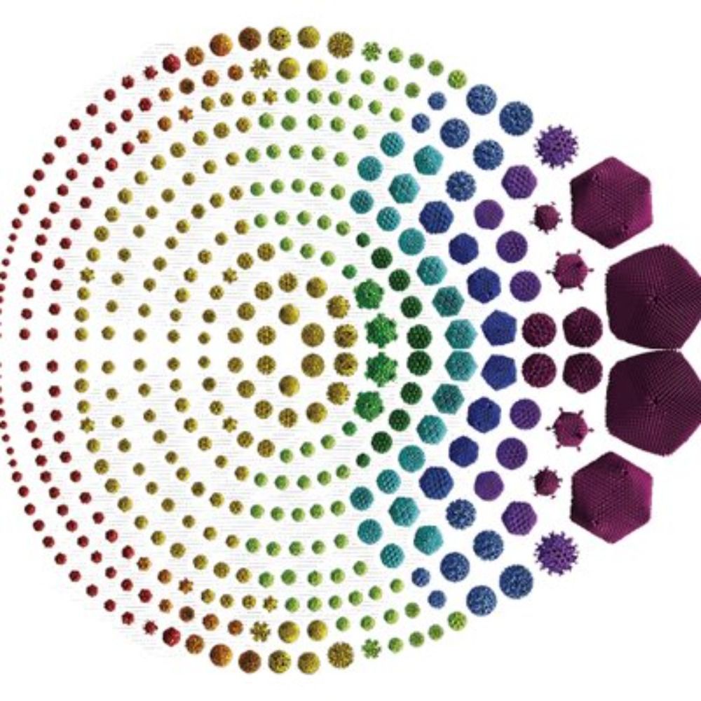
Check out our latest paper on mirusviruses, one of the most remarkable new groups of protist viruses - extremely diverse, carry lots of spliceosomal introns (including new homing introns) and are at the evolutionary crossroads between tailed phages and herpesviruses! www.nature.com/articles/s41...

Widespread and intron-rich mirusviruses are predicted to reproduce in nuclei of unicellular eukaryotes - Nature Microbiology
Environmental metagenomic explorations show that Mirusviricota lineages lack essential replication and transcription genes and contain spliceosomal introns, suggesting nuclear reproduction.
www.nature.com
November 28, 2025 at 4:43 PM
Check out our latest paper on mirusviruses, one of the most remarkable new groups of protist viruses - extremely diverse, carry lots of spliceosomal introns (including new homing introns) and are at the evolutionary crossroads between tailed phages and herpesviruses! www.nature.com/articles/s41...
Reposted by Lauren Queiss

#NatMicroPicks
A new protist with remnants of ancient mitochondrial DNA 🧬🦠
The eukaryotic tree of life grows with the addition Solarion arienae - a unique and peculiar protist species
#MicroSky
www.nature.com/articles/s41...
A new protist with remnants of ancient mitochondrial DNA 🧬🦠
The eukaryotic tree of life grows with the addition Solarion arienae - a unique and peculiar protist species
#MicroSky
www.nature.com/articles/s41...

Rare microbial relict sheds light on an ancient eukaryotic supergroup - Nature
The discovery of an unusual protist named Solarion arienae, which has a mitochondrial genome with some intriguing features, provides insight into the early radiation of eukaryotic groups.
www.nature.com
November 28, 2025 at 1:49 PM
#NatMicroPicks
A new protist with remnants of ancient mitochondrial DNA 🧬🦠
The eukaryotic tree of life grows with the addition Solarion arienae - a unique and peculiar protist species
#MicroSky
www.nature.com/articles/s41...
A new protist with remnants of ancient mitochondrial DNA 🧬🦠
The eukaryotic tree of life grows with the addition Solarion arienae - a unique and peculiar protist species
#MicroSky
www.nature.com/articles/s41...
Reposted by Lauren Queiss

How do cells adapt morphology to function? In a 🔥 preprint by @zjmaggiexu.bsky.social , with @dudinlab.bsky.social and @amyweeks.bsky.social , we identify a self-organizing single-cell morphology circuit that optimizes the feeding trap structure of the suctorian P. collini. 🧵 tinyurl.com/4k8nv926
November 18, 2025 at 4:15 PM
How do cells adapt morphology to function? In a 🔥 preprint by @zjmaggiexu.bsky.social , with @dudinlab.bsky.social and @amyweeks.bsky.social , we identify a self-organizing single-cell morphology circuit that optimizes the feeding trap structure of the suctorian P. collini. 🧵 tinyurl.com/4k8nv926
Reposted by Lauren Queiss

our next online seminar, this time with Anna Sophia Feix, on parasite-derived EVs in infection and immunity 😎 Please register here: donau-uni.zoom.us/meeting/regi...

November 6, 2025 at 5:54 PM
our next online seminar, this time with Anna Sophia Feix, on parasite-derived EVs in infection and immunity 😎 Please register here: donau-uni.zoom.us/meeting/regi...
Reposted by Lauren Queiss

So a big day for #microeukaryote #imaging. To finish "en beaute" a small video to share the behind the scenes of this first work. Such an exciting time to be a #marine #microbiologist !!
October 31, 2025 at 3:27 PM
So a big day for #microeukaryote #imaging. To finish "en beaute" a small video to share the behind the scenes of this first work. Such an exciting time to be a #marine #microbiologist !!
Reposted by Lauren Queiss

Congratulations @lqueiss.bsky.social 👏🎉😀
Lauren won the Best Poster Award at the Women in Electron Microscopy (#WeM) – Exchange and Networking event @fz-juelich.de
Pic: FZJülich/Kurt Steinhausen
www.mpi-bremen.de/en/Lauren-Qu...
#WomeninSTEM #marinescience #MachMINT @maxplanck.de #FemaleScience
Lauren won the Best Poster Award at the Women in Electron Microscopy (#WeM) – Exchange and Networking event @fz-juelich.de
Pic: FZJülich/Kurt Steinhausen
www.mpi-bremen.de/en/Lauren-Qu...
#WomeninSTEM #marinescience #MachMINT @maxplanck.de #FemaleScience

October 27, 2025 at 7:59 AM
Congratulations @lqueiss.bsky.social 👏🎉😀
Lauren won the Best Poster Award at the Women in Electron Microscopy (#WeM) – Exchange and Networking event @fz-juelich.de
Pic: FZJülich/Kurt Steinhausen
www.mpi-bremen.de/en/Lauren-Qu...
#WomeninSTEM #marinescience #MachMINT @maxplanck.de #FemaleScience
Lauren won the Best Poster Award at the Women in Electron Microscopy (#WeM) – Exchange and Networking event @fz-juelich.de
Pic: FZJülich/Kurt Steinhausen
www.mpi-bremen.de/en/Lauren-Qu...
#WomeninSTEM #marinescience #MachMINT @maxplanck.de #FemaleScience
Reposted by Lauren Queiss
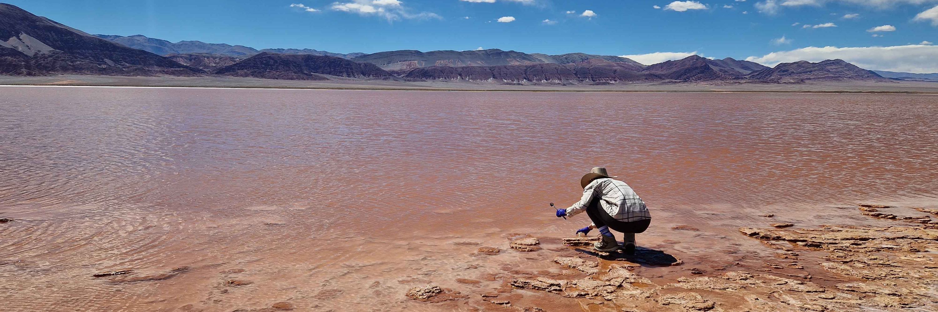
Some years ago, we discovered a modern microbialite reef under conditions resembling primitive Earth 🌋
Now we show how seasonal extremes drive microbial shifts and mineralisation, offering a window into processes that shaped Earth’s first biostructures 🪨
www.nature.com/articles/s43247-025-02764-6
Now we show how seasonal extremes drive microbial shifts and mineralisation, offering a window into processes that shaped Earth’s first biostructures 🪨
www.nature.com/articles/s43247-025-02764-6
September 24, 2025 at 1:12 PM
Some years ago, we discovered a modern microbialite reef under conditions resembling primitive Earth 🌋
Now we show how seasonal extremes drive microbial shifts and mineralisation, offering a window into processes that shaped Earth’s first biostructures 🪨
www.nature.com/articles/s43247-025-02764-6
Now we show how seasonal extremes drive microbial shifts and mineralisation, offering a window into processes that shaped Earth’s first biostructures 🪨
www.nature.com/articles/s43247-025-02764-6
Reposted by Lauren Queiss

🚨Our paper is out! 🥳
Hijacking a bacterial ABC transporter for efficient genetic code expansion.
Many congrats to everyone involved - a multi-year effort led by @taruniype.bsky.social @maxfottner.bsky.social
www.nature.com/articles/s41...
it all started years ago with a failed experiment
🧵👇 1/9
Hijacking a bacterial ABC transporter for efficient genetic code expansion.
Many congrats to everyone involved - a multi-year effort led by @taruniype.bsky.social @maxfottner.bsky.social
www.nature.com/articles/s41...
it all started years ago with a failed experiment
🧵👇 1/9

Hijacking a bacterial ABC transporter for genetic code expansion - Nature
Bacterial ATP-binding cassette (ABC) transporters can be utilized and engineered to transport non-canonical amino acids into Escherichia coli for highly efficient synthesis of proteins with novel func...
www.nature.com
October 16, 2025 at 2:06 PM
🚨Our paper is out! 🥳
Hijacking a bacterial ABC transporter for efficient genetic code expansion.
Many congrats to everyone involved - a multi-year effort led by @taruniype.bsky.social @maxfottner.bsky.social
www.nature.com/articles/s41...
it all started years ago with a failed experiment
🧵👇 1/9
Hijacking a bacterial ABC transporter for efficient genetic code expansion.
Many congrats to everyone involved - a multi-year effort led by @taruniype.bsky.social @maxfottner.bsky.social
www.nature.com/articles/s41...
it all started years ago with a failed experiment
🧵👇 1/9
Reposted by Lauren Queiss
Phages evolve fast, or do they?
In oysters, some stay identical for years.
With >1,200 phages & 600 Vibrio genomes, we reveal long-term stability and new mobile elements.
Proud of this collaborative work across our teams (Roscoff-UdeM and @epcrocha.bsky.social www.biorxiv.org/cgi/content/...
In oysters, some stay identical for years.
With >1,200 phages & 600 Vibrio genomes, we reveal long-term stability and new mobile elements.
Proud of this collaborative work across our teams (Roscoff-UdeM and @epcrocha.bsky.social www.biorxiv.org/cgi/content/...

Ecological constraints foster both extreme viral-host lineage stability and mobile element diversity in a marine community
Phages are typically viewed as very rapidly evolving biological entities. Little is known, however, about whether and how phages can establish long-term genetic stability. We addressed this eco-evolut...
www.biorxiv.org
October 12, 2025 at 9:16 PM
Phages evolve fast, or do they?
In oysters, some stay identical for years.
With >1,200 phages & 600 Vibrio genomes, we reveal long-term stability and new mobile elements.
Proud of this collaborative work across our teams (Roscoff-UdeM and @epcrocha.bsky.social www.biorxiv.org/cgi/content/...
In oysters, some stay identical for years.
With >1,200 phages & 600 Vibrio genomes, we reveal long-term stability and new mobile elements.
Proud of this collaborative work across our teams (Roscoff-UdeM and @epcrocha.bsky.social www.biorxiv.org/cgi/content/...
Reposted by Lauren Queiss

Balancing stability and flexibility when reshaping archaeal membranes.
buff.ly/4yqUdBj
buff.ly/4yqUdBj
October 10, 2025 at 8:03 PM
Balancing stability and flexibility when reshaping archaeal membranes.
buff.ly/4yqUdBj
buff.ly/4yqUdBj
Reposted by Lauren Queiss

Oceanic cyanobacterial photosynthesis is negatively affected by viral NblA proteins www.biorxiv.org/content/10.1...


October 12, 2025 at 8:26 AM
Oceanic cyanobacterial photosynthesis is negatively affected by viral NblA proteins www.biorxiv.org/content/10.1...
Reposted by Lauren Queiss

Compositional analysis of bacterial peptidoglycan: insights from peptidoglycomics into structure and function | Journal of Bacteriology https://journals.asm.org/doi/full/10.1128/jb.00359-25?af=R

Compositional analysis of bacterial peptidoglycan: insights from peptidoglycomics into structure and function | Journal of Bacteriology
Peptidoglycan (PG) is a crucial biopolymer in the bacterial cell wall that has been the subject of intense study since it was first isolated in the early 1950s (1, 2). Over the last 70 years, extensive research has expanded our understanding of the structure and function of this microbial biopolymer. Recent advances in mass spectrometry and bioinformatics have revolutionized PG analysis, enabling a comprehensive detection of individual components and their global composition within bacterial cells. Like genomics, transcriptomics, and proteomics, peptidoglycomics is the non-targeted, non-biased detection of all elements that comprise the overall PG structure. Peptidoglycomic analyses can identify and monitor hundreds of potential compositional changes that occur within the PG structure of a cell. By comparison, traditional methods of analyzing PG composition only distinguish a relatively limited number of PG components. Therefore, peptidoglycomic approaches produce a detailed global overview of the PG structural elements and give unprecedented insight into the physiological function of this biopolymer within the bacterial cell.
journals.asm.org
October 11, 2025 at 1:41 PM
Compositional analysis of bacterial peptidoglycan: insights from peptidoglycomics into structure and function | Journal of Bacteriology https://journals.asm.org/doi/full/10.1128/jb.00359-25?af=R
Reposted by Lauren Queiss

Marine particles harbor microbial communities. However, to study them, they must be separated from the particle. How? This #AppEnvMicro article outlines an optimized method using detergents to dissociate microbes from marine particles. Get the details: asm.social/2DG

October 10, 2025 at 4:43 PM
Marine particles harbor microbial communities. However, to study them, they must be separated from the particle. How? This #AppEnvMicro article outlines an optimized method using detergents to dissociate microbes from marine particles. Get the details: asm.social/2DG
Reposted by Lauren Queiss

No Genetics? Try ASOs – A non-genetic approach to silence genes at the phage-host interface. We use it to study jumbo phage biology and anti-phage defence.
@jorg-vogel-lab.bsky.social @helmholtz-hiri.bsky.social
@uni-wuerzburg.de @helmholtzhzi.bsky.social
published now in @nature.com
@jorg-vogel-lab.bsky.social @helmholtz-hiri.bsky.social
@uni-wuerzburg.de @helmholtzhzi.bsky.social
published now in @nature.com

Programmable antisense oligomers for phage functional genomics - Nature
Establishing antisense oligomers as versatile, non-genetic tools to silence phage mRNAs opens applications in basic research and biotechnology, as shown by identifying essential factors for propagatio...
www.nature.com
September 11, 2025 at 9:10 AM
No Genetics? Try ASOs – A non-genetic approach to silence genes at the phage-host interface. We use it to study jumbo phage biology and anti-phage defence.
@jorg-vogel-lab.bsky.social @helmholtz-hiri.bsky.social
@uni-wuerzburg.de @helmholtzhzi.bsky.social
published now in @nature.com
@jorg-vogel-lab.bsky.social @helmholtz-hiri.bsky.social
@uni-wuerzburg.de @helmholtzhzi.bsky.social
published now in @nature.com
Reposted by Lauren Queiss

Her story celebrates women in marine science and the fight to protect our blue planet — all from a small, storm-tossed island in the Irish Sea.
If you care about the ocean’s future, this one’s a tide worth catching. 🌊✨
📘 Spring Tides by Fiona Gell
🔹 Reviewed by Matthew Bunce FMBA
#MarineScience
If you care about the ocean’s future, this one’s a tide worth catching. 🌊✨
📘 Spring Tides by Fiona Gell
🔹 Reviewed by Matthew Bunce FMBA
#MarineScience
October 9, 2025 at 3:03 PM
Her story celebrates women in marine science and the fight to protect our blue planet — all from a small, storm-tossed island in the Irish Sea.
If you care about the ocean’s future, this one’s a tide worth catching. 🌊✨
📘 Spring Tides by Fiona Gell
🔹 Reviewed by Matthew Bunce FMBA
#MarineScience
If you care about the ocean’s future, this one’s a tide worth catching. 🌊✨
📘 Spring Tides by Fiona Gell
🔹 Reviewed by Matthew Bunce FMBA
#MarineScience
Reposted by Lauren Queiss
Book spotlight - SPRING TIDES by Fiona Gell
Book Spotlight: Spring Tides by Fiona Gell
A lyrical dive into the Isle of Man’s marine life — and what it teaches us about ocean conservation, policy, and the human heart.
Book Spotlight: Spring Tides by Fiona Gell
A lyrical dive into the Isle of Man’s marine life — and what it teaches us about ocean conservation, policy, and the human heart.


October 9, 2025 at 3:03 PM
Book spotlight - SPRING TIDES by Fiona Gell
Book Spotlight: Spring Tides by Fiona Gell
A lyrical dive into the Isle of Man’s marine life — and what it teaches us about ocean conservation, policy, and the human heart.
Book Spotlight: Spring Tides by Fiona Gell
A lyrical dive into the Isle of Man’s marine life — and what it teaches us about ocean conservation, policy, and the human heart.
I’m excited to take part in the very first Women in Electron Microscopy (WEM) Conference hosted by @fz-juelich.de for breaking barriers and building networks!
#womeninSTEM #ElectronMicroscopy
#womeninSTEM #ElectronMicroscopy

October 8, 2025 at 8:16 PM
I’m excited to take part in the very first Women in Electron Microscopy (WEM) Conference hosted by @fz-juelich.de for breaking barriers and building networks!
#womeninSTEM #ElectronMicroscopy
#womeninSTEM #ElectronMicroscopy
Reposted by Lauren Queiss
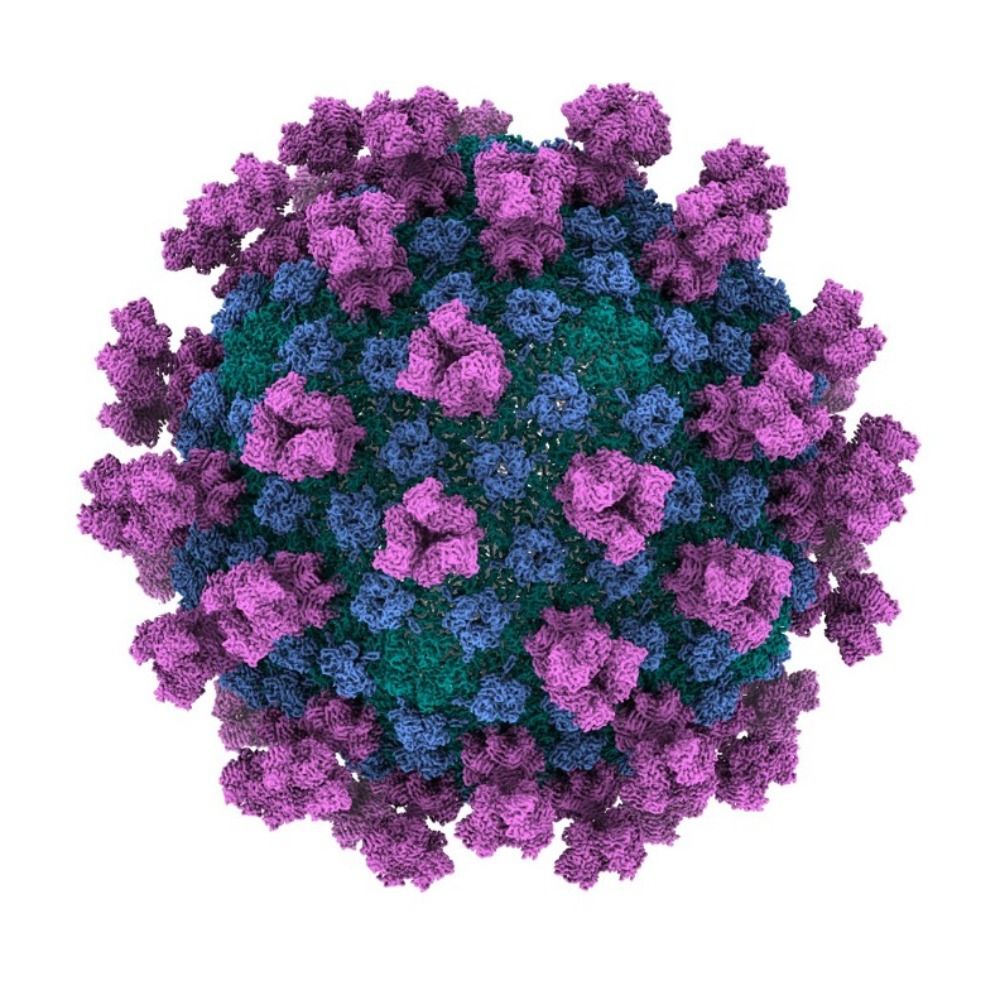
Out in Science Advances: Our #cryoEM structure of HFTV1, a virus infecting the halophile #archaea. *First full atomic structure (containing all structural proteins) of any tailed virus!* Congrats and thanks to all co-authors and our fantastic collaborators! www.science.org/doi/10.1126/...

Cryo-EM resolves the structure of the archaeal dsDNA virus HFTV1 from head to tail
This structure of an archaeal tailed virus (arTV) provides detailed insights into arTV assembly and infection mechanisms.
www.science.org
October 6, 2025 at 11:31 AM
Out in Science Advances: Our #cryoEM structure of HFTV1, a virus infecting the halophile #archaea. *First full atomic structure (containing all structural proteins) of any tailed virus!* Congrats and thanks to all co-authors and our fantastic collaborators! www.science.org/doi/10.1126/...
Reposted by Lauren Queiss

Jane Goodall, known for her pioneering work with chimpanzees, has passed away aged 91
go.nature.com/46K10ja
go.nature.com/46K10ja

Jane Goodall’s legacy: three ways she changed science
The primatologist challenged what it meant to be a scientist.
go.nature.com
October 2, 2025 at 9:38 AM
Jane Goodall, known for her pioneering work with chimpanzees, has passed away aged 91
go.nature.com/46K10ja
go.nature.com/46K10ja
Reposted by Lauren Queiss

then turned that knowledge into a global movement for conservation. Her work made it impossible to separate what we learn from our obligation to protect it. Generations of researchers have tried to follow that example." 2/2

October 2, 2025 at 4:51 AM
then turned that knowledge into a global movement for conservation. Her work made it impossible to separate what we learn from our obligation to protect it. Generations of researchers have tried to follow that example." 2/2
Reposted by Lauren Queiss
From IGI Founder Jennifer Doudna: "Jane Goodall showed us what a life in science could look like: rigorous discovery paired with fierce advocacy for what you study. She gave the world six decades of groundbreaking research on chimpanzees and their habitats... 1/2

October 2, 2025 at 4:51 AM
From IGI Founder Jennifer Doudna: "Jane Goodall showed us what a life in science could look like: rigorous discovery paired with fierce advocacy for what you study. She gave the world six decades of groundbreaking research on chimpanzees and their habitats... 1/2
Reposted by Lauren Queiss
We are excited to share our preprint describing how Sulfolobus cells coordinate DNA segregation with cell division! In eukaryotes this type of regulation involves checkpoints and CDK-cyclins. But how does this work in archaea? This is the question we ask in our paper: www.biorxiv.org/content/10.1...

Temporal and spatial coordination of DNA segregation and cell division in an archaeon.
Cells must coordinate DNA segregation with cytokinesis to ensure that each daughter cell inherits a complete genome. Here, we explore how DNA segregation and division are mechanistically coupled in ar...
www.biorxiv.org
May 29, 2025 at 3:36 PM
We are excited to share our preprint describing how Sulfolobus cells coordinate DNA segregation with cell division! In eukaryotes this type of regulation involves checkpoints and CDK-cyclins. But how does this work in archaea? This is the question we ask in our paper: www.biorxiv.org/content/10.1...