Colin Nichols Lab
@colinnicholslab.bsky.social
290 followers 620 following 38 posts
Posting from Colin Nichols' electrophysiology lab at WashU. Focused on ion channels biophysics & role in physiology and pathology.
Posts Media Videos Starter Packs
Reposted by Colin Nichols Lab
colinnicholslab.bsky.social
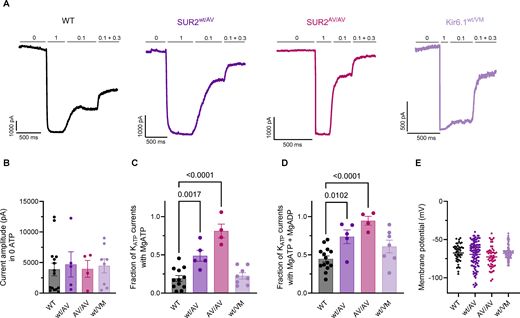
These results shed light on the pathophysiologic relevance of SUR2-dependent KATP channel subunits in skeletal muscle and highlight their role in fatiguing conditions. (6/n)
colinnicholslab.bsky.social
These effects could be prevented in the presence of the KATP channel inhibitor glibenclamide, indicating that the increased fatigue of isolated muscles is a direct consequence of overactive sarcolemmal KATP channels. (5/n)
colinnicholslab.bsky.social
Ex vivo testing of isolated SUR2[A478V], but not Kir6.1[V65M], muscles showed an early onset of fatigue and a marked intra-tetanic decline of force. (4/n)
colinnicholslab.bsky.social
Direct consequences of CS mutations on sarcolemma KATP channels on muscle contractility are currently unclear. Here, we assessed contractility in isolated fast- and slow-twitch muscles from two mouse models of CS, carrying GOF mutations Kir6.1[V65M] or SUR2[A478V]. (3/n)
colinnicholslab.bsky.social
Cantu syndrome (CS) is a rare disease caused by gain-of-function (GOF) mutations of Kir6.1 or SUR2 subunits of ATP-sensitive potassium (KATP) channels. CS patients display a constellation of symptoms, including muscle weakness and fatigue. (2/n)
colinnicholslab.bsky.social
Check out our new publication "Muscle fatigue arising intrinsically from SUR2- but not Kir6.1-dependent gain-of-function in Cantu syndrome mice" 🧪 @rosca26.bsky.social
A 🧵 1/n
jgp.org
Scala et al. @colinnicholslab.bsky.social assess #SkeletalMuscle properties in gain-of-function knock-in mouse models of Cantu Syndrome. Isolated SUR2 GOF, but not Kir6.1 GOF muscles showed enhanced fatiguing that was reversed by the KATP channel inhibitor glibenclamide.
Muscle fatigue arising intrinsically from SUR2- but not Kir6.1-dependent gain-of-function in Cantu syndrome mice
We assessed skeletal muscle properties in GOF knock-in mouse models of Cantu Syndrome. In isolated myofibers there was enhanced Mg-nucleotide activation in
rupress.org
Reposted by Colin Nichols Lab
rupress.org
New in @jgp.org: Zangerl-Plessl, Lee, et al. utilized MD simulations to reveal that PIP2 potentiated clockwise twisting motions in individual Kir2 #IonChannel cytoplasmic subunits, as well as concerted dynamics among the four subunits. rupress.org/jgp/article/...
@colinnicholslab.bsky.social
colinnicholslab.bsky.social
These motions are reduced when PIP2 is removed, leading to narrowing of the critical gate at the M2 helix bundle crossing (HBC), but expansion at the region G-loop, as well as reduced overall fourfold symmetry, in turn coupled to cessation of ion permeation. (5/n)
colinnicholslab.bsky.social
We have carried out full atomistic MD simulations, which indicate PIP2-dependent conformational changes that are coupled to opening and closing of the channel. In the presence of bound PIP2, the cytoplasmic domain performs clockwise twisting motions. (4/n)
colinnicholslab.bsky.social
Most Kir2 channel structures determined in complex with PIP2 molecules are in a closed state, requiring additional conformational changes for channel opening. (3/n)
colinnicholslab.bsky.social
Inwardly rectifying potassium (Kir) channel activity is important in the control of membrane potentials and is regulated through various ligands, including Phosphatidyl-4,5-bisphosphate (PIP2)(2/n)
colinnicholslab.bsky.social
Check out our new publication "PIP2-driven cytoplasmic domain motions are coupled to Kir2 channel gating" 🧪
A 🧵 1/n
jgp.org
Eva-Maria Zangerl-Plessl, Sun-Joo Lee, et al. utilized MD simulations to reveal that PIP2 potentiated clockwise twisting motions in individual Kir2 #IonChannel cytoplasmic subunits, as well as concerted dynamics among the four subunits. rupress.org/jgp/article/...
@colinnicholslab.bsky.social
colinnicholslab.bsky.social
We love it Stephen, thank you! 🧪
stevestonedraws.bsky.social
Dr. Colin G. Nichols shared how the KATP channel drives both neonatal diabetes & congenital hyperinsulinism. 🧬 Key insights: SUR1 mutations, shifts in calcium sensitivity, & why diazoxide works (when sulfonylureas may not).
@washumedicine.bsky.social @colinnicholslab.bsky.social #EndoSky
colinnicholslab.bsky.social
Our data provide definitive support for a paradoxical form of MODY associated with KATP channel LOF that is genetically and mechanistically distinct from a late diagnosis of neonatal diabetes resulting from KATP GOF. (5/n)
colinnicholslab.bsky.social
In contrast to the naïve prediction that diabetes should be associated with KATP gain-of-function (GOF, as in KATP-dependent neonatal diabetes), each mutation caused mild to severe loss-of-function (LOF), through distinct molecular mechanisms. (4/n)
colinnicholslab.bsky.social
We report genotype-phenotype information from a set of patients clinically diagnosed with maturity-onset diabetes of the young (MODY) and carrying coding variants in the KATP regulatory subunit gene ABCC8. (3/n)
colinnicholslab.bsky.social
Pancreatic β-cell ATP-sensitive K+ (KATP) channel closure underlies electrical excitability and insulin release, but loss or inhibition of KATP channels can lead to paradoxical crossover from hyperinsulinism plus hypoglycemia, to glucose intolerance or diabetes. (2/n)
colinnicholslab.bsky.social
Join us today for a new exciting CIMED seminar! 🧪 @osamaharraz.bsky.social
colinnicholslab.bsky.social
Join us today for a new exciting CIMED seminar! 🧪
colinnicholslab.bsky.social
🧪
cryoempapers.bsky.social
Dynein light chains 1 and 2 are auxiliary proteins of pH-sensitive Kir4.1 channels pubmed.ncbi.nlm.nih.gov/40074079/ #cryoem