Danielle Spitzer
@dspitzer.bsky.social
170 followers
160 following
23 posts
🧬👩🏻🏫 Teaching Assistant Professor at Pitt BioSci ⭐️💙 Biology nerd enthusiastic about evidence-based and inclusive pedagogy. She/her.
Posts
Media
Videos
Starter Packs
Pinned

Reposted by Danielle Spitzer

Reposted by Danielle Spitzer
Reposted by Danielle Spitzer


Reposted by Danielle Spitzer
Reposted by Danielle Spitzer


Reposted by Danielle Spitzer


Reposted by Danielle Spitzer
Eric 🏳️🌈
@erichastie.bsky.social
· Feb 27
Teaching Asst or Teaching Associate Professor
The position calls for teaching three classes per semester, which may include fundamental classes for biology majors such as cell biology and genetics, and biodiversity (ecology, evolution, and organi...
unc.peopleadmin.com

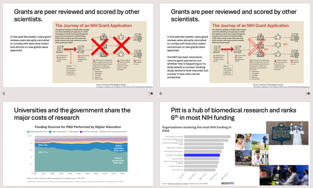
![Slide 12: Reality: NIH consistently spends about 27% of grant money on indirect costs. Graph showing costs for tope 30 universities are all approx. at 27%
Slide 13: Major, abrupt funding cuts were announced and blocked within the last week. The policy announced last Friday would cap reimbursement to universities for indirect costs at 15%. At least as of yesterday, this has been temporarily halted. Screenshots of news articles: "New NIH policy: Pitt faces big cut in federal research money[Pittsburgh Post-Gazette" "‘Science is non-partisan’: Pitt researchers face NIH funding freeze [THEPITTNEWS]" "Judge freezes NIH funding cuts nationwide as Pittsburgh universities join legal challenge [90.5 WESA]"
Slides 14/15. Long emails from Pitt officials (chancellor, vice chancellors, etc.) stressing things like "much is at stake with the proposed cuts to indirect costs...A significant reduction of these funds will result in irreparable harm for University operations"](https://cdn.bsky.app/img/feed_thumbnail/plain/did:plc:n63e5qt7nsa4ctd32ados5u4/bafkreic2g7wdibzn4jcuhkqtrae2wanyyub3esnbwqfplpkhbsz7jjybci@jpeg)


Danielle Spitzer
@dspitzer.bsky.social
· Dec 22