
Eva-Maria Schentarra
@evaschentarra.bsky.social
56 followers
77 following
12 posts
Biotechnologist - PhD student @jungmannlab.bsky.social
Enjoying the little things in life using #DNAPAINT 🔬🧬
@mpibiochem.bsky.social
@lmumuenchen.bsky.social
@imprs-ml.bsky.social
Posts
Media
Videos
Starter Packs

Reposted by Eva-Maria Schentarra


Reposted by Eva-Maria Schentarra

Reposted by Eva-Maria Schentarra


Reposted by Eva-Maria Schentarra

Reposted by Eva-Maria Schentarra

JungmannLab
@jungmannlab.bsky.social
· May 7
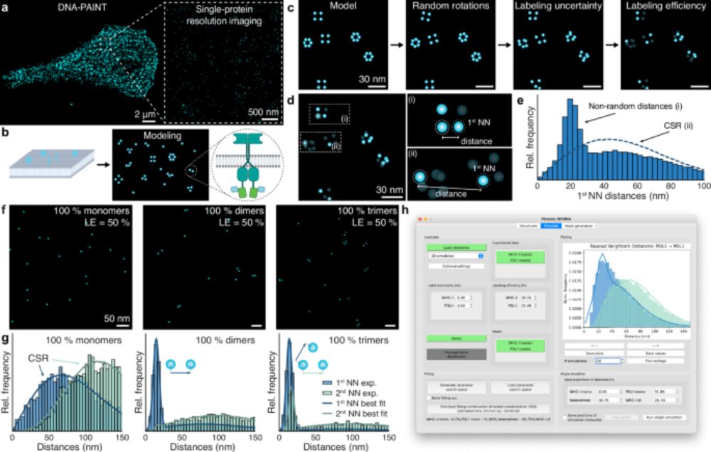
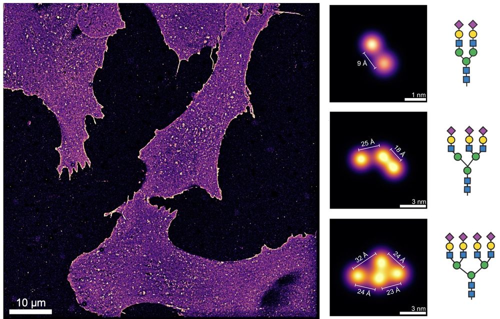
Spatial and stoichiometric in situ analysis of biomolecular oligomerization at single-protein resolution - Nature Communications
Extracting quantitative information on biomolecular oligomerisation with high resolution remains a significant challenge. Here, the authors propose SPINNA, a framework that compares nearest-neighbour ...
www.nature.com
Reposted by Eva-Maria Schentarra
JungmannLab
@jungmannlab.bsky.social
· May 7
Spatial and stoichiometric in situ analysis of biomolecular oligomerization at single-protein resolution - Nature Communications
Extracting quantitative information on biomolecular oligomerisation with high resolution remains a significant challenge. Here, the authors propose SPINNA, a framework that compares nearest-neighbour ...
www.nature.com
Reposted by Eva-Maria Schentarra

Eugenio F. Fornasiero
@forna.bsky.social
· Apr 22

Nanobinders for Synaptotagmin 1 enable the analysis of synaptic vesicle dynamics in rodent and human models.
Synaptic neurotransmission is a critical hallmark of brain activity and one of the first processes to be affected in neural diseases. Monitoring this process, and in particular synaptic vesicle recycl...
www.biorxiv.org
Reposted by Eva-Maria Schentarra


Reposted by Eva-Maria Schentarra

Malina Iwanski
@mkiwanski.bsky.social
· Feb 9

Polarity reversal of stable microtubules during neuronal development
Neurons critically depend on long-distance transport orchestrated by motor proteins walking over their highly asymmetric microtubule cytoskeleton. These microtubules are organized uniformly in axons w...
www.biorxiv.org
Reposted by Eva-Maria Schentarra