Kevin Staras
@kevinstaras.bsky.social
280 followers
220 following
24 posts
NeuroProf at University of Sussex, UK • Synapses, Circuits, Plasticity, Disease, Decision-making
https://www.thestaraslab.org/
Posts
Media
Videos
Starter Packs
Reposted by Kevin Staras

Reposted by Kevin Staras

Matteo Carandini
@carandinilab.net
· Jun 17
Reposted by Kevin Staras

Reposted by Kevin Staras

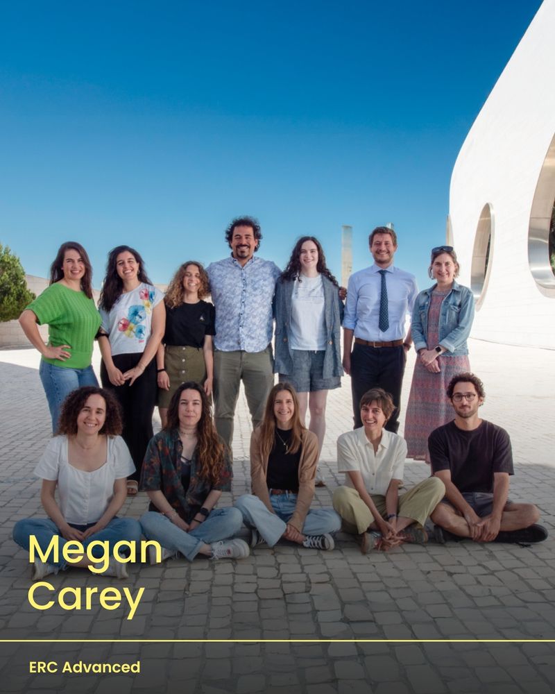
Megan Carey
@megancarey.bsky.social
· Jun 17
Reposted by Kevin Staras
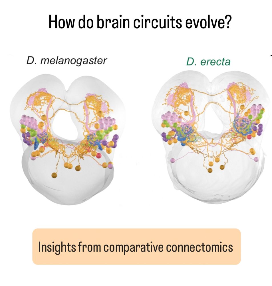
Megan Carey
@megancarey.bsky.social
· Jun 17
Reposted by Kevin Staras


Reposted by Kevin Staras


Reposted by Kevin Staras

Michael Reiser
@michaelreiser.bsky.social
· Mar 27
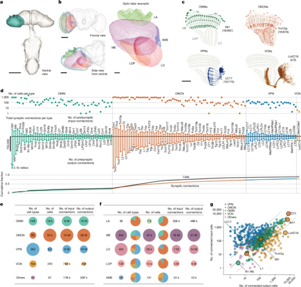
Connectome-driven neural inventory of a complete visual system - Nature
A connectome of the right optic lobe from a male fruitfly is presented together with an extensive collection of genetic drivers matched to a comprehensive neuron-type catalogue.
www.nature.com